Can a chest X-ray flag osteoporosis?
A peer-reviewed Korean study externally validated an AI prescreener in three cohorts totalling 153,058 people. Its simulated workflow preserved most osteoporosis detections while halving DXA use, but it has not yet proved benefit in prospective care.
Editorial responsibility: The Impact of AI Editorial Desk · Report a factual concern
At a glance
- 1The model was developed from 46,478 paired chest-radiograph and DXA examinations belonging to 26,757 patients, then tested in three independent cohorts containing 6,391, 24,132 and 122,535 people.
- 2Reported AUROCs for osteoporosis were 0.88 in the temporally separated hospital cohort and 0.93 and 0.92 in the two health-check cohorts.
- 3A simulation suggested that model-based prescreening could halve DXA use while retaining 98–99% of osteoporosis cases, but no prospective trial tested referrals, treatment, fractures or patient outcomes.
Research topic
Opportunistic osteoporosis prescreening from routine chest radiographs
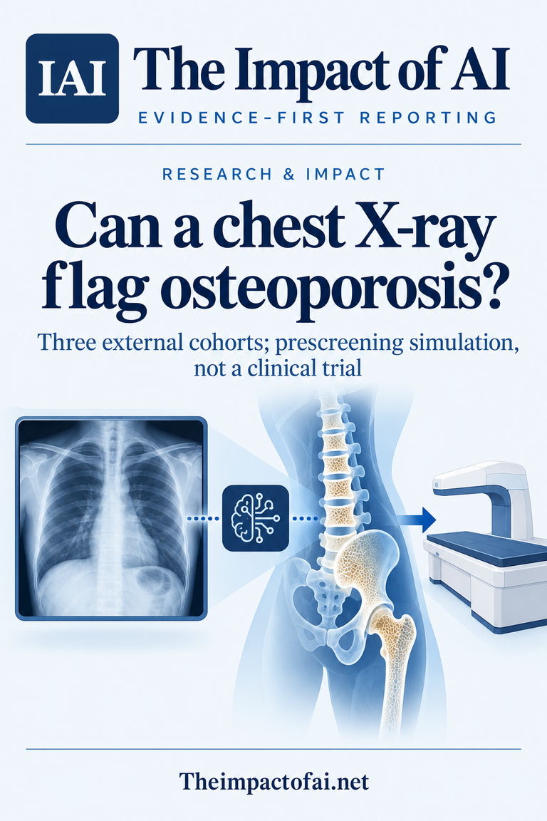
The direct answer: useful as a prescreener, not a diagnosis
A routine chest X-ray appears capable of helping decide who should receive the definitive bone-density test, at least in the Korean populations examined. The new model, AI-OsRM, separated people with and without osteoporosis with an area under the receiver operating characteristic curve of 0.88 in a temporally separated hospital cohort and 0.93 and 0.92 in two health-check cohorts. Those are strong discrimination results across three independent test populations.
They do not mean a chest radiograph can replace dual-energy X-ray absorptiometry, or DXA. The study treats the AI output as a prescreening signal: a way to identify more people who may merit DXA from imaging already acquired for another reason. The study was retrospective, and the claimed reduction in DXA use came from a simulation. It did not observe clinicians acting on alerts or measure diagnoses, treatment, fractures, costs or harms in prospective care.[1]
The development set linked images to the reference test
The researchers developed AI-OsRM using 46,478 paired chest radiograph and DXA examinations from 26,757 patients at a tertiary referral hospital between 2016 and 2022. DXA supplies the reference measurement of bone mineral density used in osteoporosis care. Pairing it with a chest image lets the model learn image patterns associated with osteoporosis and with reduced bone mineral density, rather than relying on radiologist impressions alone.
The architecture was based on a residual neural network and used masked-autoencoder pretraining. That design detail matters less clinically than the data path: the model learned from people who had both tests, a selected group that may differ from everyone receiving a chest X-ray. Referral patterns, age, illness and local practice can influence who reaches DXA. A robust deployment assessment must therefore examine calibration and errors in the intended screening population, not only ranking performance in retrospective cohorts.[1]
Three external cohorts supplied the study’s strongest evidence
External validation covered 153,058 people: 6,391 in a later cohort from Seoul National University Hospital, 24,132 in one health-promotion-centre cohort and 122,535 in another health-screening cohort. The hospital cohort tested temporal separation from development data, while the two much larger screening cohorts tested a different clinical context. This breadth is an important advance over a single random split from one institution.
The AUROC was 0.88, with a 95% confidence interval of 0.87 to 0.89, for both osteoporosis and reduced bone density in the hospital cohort. For osteoporosis in the health-check cohorts, it was 0.93 with a 0.92–0.93 interval and 0.92 with a 0.92–0.92 interval. AUROC describes ranking across thresholds; it does not show how many alerts would be false in a particular clinic. Positive predictive value will change with prevalence, so threshold-specific results and local calibration remain essential.[1]
The headline workflow result is simulated
The authors simulated a two-stage pathway in which AI-OsRM first filters routine chest radiographs and selected people proceed to DXA. Under the tested settings, DXA use fell by about 50% while the pathway retained 98–99% of osteoporosis cases and 80–90% of people with reduced bone mineral density. That trade-off is the most practical result because DXA capacity and attendance can constrain screening.
A simulation cannot capture every consequence of deployment. Real alerts may be ignored, duplicated or sent to people already tested; clinicians may change thresholds; patients may decline DXA; and false positives can create anxiety and extra appointments. Missed cases matter too, particularly when reduced bone density rather than established osteoporosis is the target. A prospective trial should predefine referral thresholds and count completed DXA tests, newly treated cases, downstream investigations and adverse effects.[1]
What patients and health services could gain
Osteoporosis is often silent until a fragility fracture. Chest radiographs are common, quick and already available in many records, so opportunistic analysis could identify people who otherwise would not be assessed. The potential benefit is not another scan generated for AI. It is extracting a cautious risk signal from an existing image, then using an established diagnostic test and ordinary clinical evaluation before making treatment decisions.
For health services, the promise is to target scarce DXA appointments without simply lowering screening volume. That requires measuring who is reached. A model trained and validated in South Korea may perform differently where disease prevalence, body composition, equipment, image acquisition and access to follow-up differ. If disadvantaged patients are less likely to complete DXA after an alert, technically accurate prescreening could still widen rather than close gaps in fracture prevention.[1]
How this changes the wider evidence picture
A 2026 systematic review of 57 opportunistic osteoporosis-AI studies found encouraging performance but substantial heterogeneity. Most studies were retrospective and single-centre, and only 36% included external validation. Smaller and single-centre studies tended to report higher accuracy. Against that background, three independent cohorts—including two large health-check populations—make this paper more informative than an internal benchmark alone.
The new study does not remove the review’s central translation problem. External retrospective validation is necessary, not sufficient. Health systems still need prospective evaluation, transparent reporting of thresholds and subgroup performance, and a clear role for the tool as an adjunct rather than a diagnosis. The most important comparator is not another algorithm’s AUROC but the real screening pathway used today: who receives DXA, who is missed and what extra burden a new alert creates.[1][2]
Funding, interests and what would change the assessment
The work was supported by Korean government National Research Foundation grants and the Seoul National University Bundang Hospital Research Fund. The paper states that the other authors declared no competing financial or non-financial interests. Public funding and peer review strengthen transparency, but they do not independently validate a model built within the same clinical research ecosystem that supplied much of the evidence.
Confidence would rise with a registered prospective multi-centre study outside South Korea, using frozen software and thresholds before the first patient is enrolled. It should report sensitivity, specificity, predictive values and calibration by age, sex, ethnicity, site and equipment, then follow completed referrals, diagnoses, treatment and fractures. Until then, this is credible evidence that a chest-X-ray model may improve case-finding efficiency—not evidence that it safely replaces DXA or prevents fractures.[1]
What this means for people
- People should not interpret a chest X-ray or an AI score as an osteoporosis diagnosis; DXA and clinical assessment remain necessary.
- Opportunistic prescreening could reach people before a first fracture without requiring an additional image solely for AI analysis.
- Health services would need to ensure alerts lead to equitable follow-up rather than additional burden for patients least able to attend DXA.
Global context
The evidence comes from South Korean hospital and health-check cohorts. Countries differ in osteoporosis prevalence, screening guidance, radiography equipment, DXA capacity and access to follow-up. A scalable opportunity exists because chest X-rays are common worldwide, but safe adoption requires local calibration, privacy safeguards and proof that referrals improve care rather than merely generating more alerts.
What the evidence does not yet show
- The development and validation were retrospective, and the proposed DXA-saving pathway was simulated rather than tested in live care.
- All reported cohorts were Korean, so calibration and performance may not transfer to other populations, prevalence levels, equipment or referral pathways.
- AUROC does not by itself specify false-alert burden or predictive value at a chosen clinical threshold.
- The study did not measure completed referrals, treatment decisions, fracture prevention, patient experience or cost-effectiveness.
- A prescreening result is not a diagnosis and should not replace DXA or clinical assessment.
What to watch next
- Prospectively registered, multi-centre trials with frozen thresholds.
- Calibration and predictive values in populations outside South Korea.
- Subgroup performance by age, sex, ethnicity, equipment and care setting.
- Completed DXA referrals, false-alert workload, treatment and fracture outcomes.
- Independent comparison with existing age- and risk-based referral pathways.
Living evidence record
Impact record IAI-0Z30DA4
Evidence stage
Studied
Confidence
Supported
Reporting basis
Source analysis
Independent or research support
Present
Record status
Monitoring
Last checked
9 October 2026
Source trail
2 direct sources across 1 source type.
People impact
Documented in this record.
Uncertainty
Limits and next checks are explicit.
Stages describe the evidence available—not whether a technology is good or bad. See the public method.
Related-source reporting disclosure
This record analyses 2 linked source records around the same underlying development. The extra records add method, date or context, but they do not by themselves constitute independent replication of every performance claim or predicted outcome.
Evidence trail
Sources used for this report
Links checked 9 October 2026
This report is labelled source analysis. We summarise and analyse source material in our own words; company statements remain attributed claims until independently supported. Translated summaries preserve the meaning of the original source and link back to it. Read our editorial standards.
Continue the story
Related reporting
Health & Life Sciences
How much does MedGemma improve medical AI?
A peer-reviewed study reports gains over similarly sized base models across medical image questions, chest X-ray classification and simulated agent tasks. The evidence comes from benchmarks and small specialist reviews, not prospective clinical deployment or patient outcomes.
9 min · 2 sources
Health & Life Sciences
Can AI improve GLP-1 and weight-loss treatment?
A new analysis of research from China and South Korea: predicting treatment response and designing a different drug candidate are promising ideas, but neither establishes that using AI improves weight-loss outcomes.
6 min · 4 sources
Health & Life Sciences
Can AI reliably predict facial growth?
A registered systematic review found 12 studies of AI-based craniofacial or mandibular growth prediction, with samples from 33 to 639 people. Seven studies were at high risk of bias and five at unclear risk; none had independent external validation.
9 min · 1 source
The Impact Brief
Keep the evidence trail, not the noise.
Get the most consequential AI developments with direct sources and clear limits.
Reader commentary
Add evidence, experience or a question
No account is required. Reader notes are published after a brief civility, relevance and safety check; disagreement is welcome.
Explore commentary across the portal →Published reader notes
0No published reader notes yet. You can start the evidence-led discussion above.
Prefer a private correction or response? Contact the newsroom.