Can AI discover new MRI signs for glioblastoma?
It generated eight candidate visual signs from 106 glioblastoma scans, but only one was externally tested. Two radiologists achieved moderate discrimination between 50 glioblastomas and 50 metastases, so this is a discovery signal—not a clinical diagnostic.
Editorial responsibility: The Impact of AI Editorial Desk · Report a factual concern
At a glance
- 1The pipeline analysed a development dataset of 106 supratentorial glioblastomas and generated eight candidate semantic imaging signs.
- 2Only the repeatedly proposed cauliflower sign received an external reader test, using 50 glioblastomas and 50 metastases assessed independently by two radiologists.
- 3Reader AUCs were 0.73 and 0.77, with overlapping uncertainty intervals; the study demonstrates a discovery workflow, not a deployable diagnostic or better patient outcomes.
Research topic
Whether an agentic AI pipeline can convert quantitative tumour-shape features into human-readable MRI signs and whether radiologists can use one proposed sign to distinguish glioblastoma from metastasis
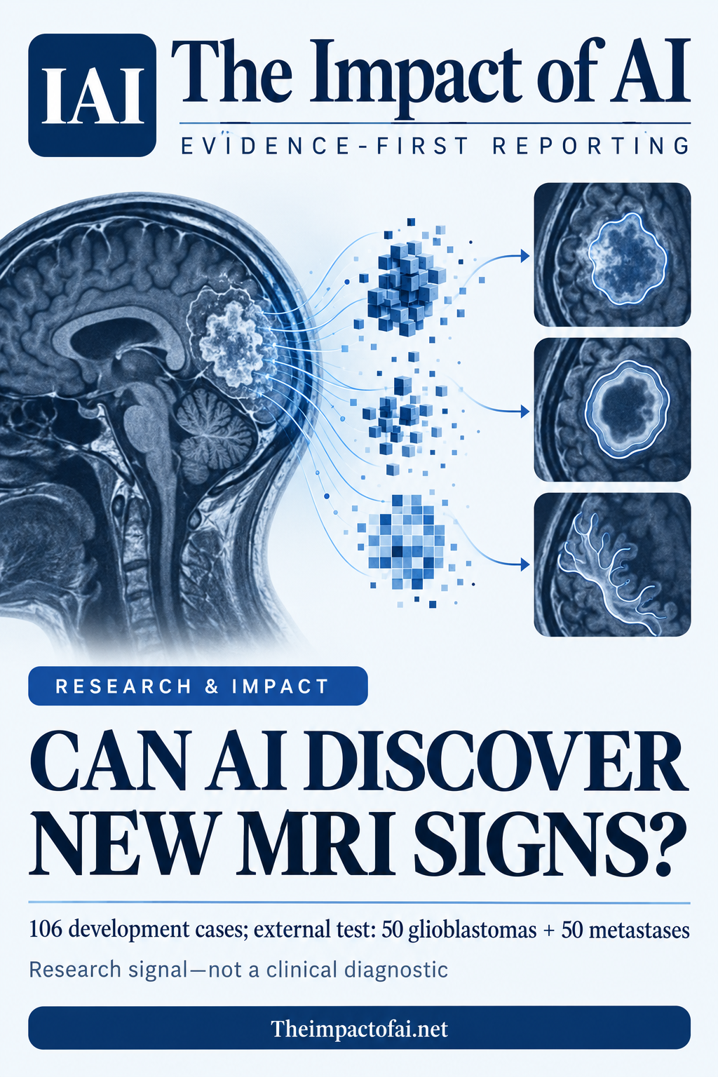
The answer is a promising discovery method, not a diagnostic
An agentic AI pipeline produced candidate MRI signs that radiologists could understand and test, and one sign showed moderate ability to separate glioblastoma from brain metastasis in an external cohort. That is meaningful because radiomics usually yields numerical features that can be difficult to translate into the visual vocabulary clinicians use. It is not evidence that an autonomous system can diagnose a brain tumour, select treatment or improve survival.
The peer-reviewed study’s strongest result concerns the proposed ‘cauliflower sign’. Two radiologists independently applied it to 100 external cases and recorded areas under the receiver operating characteristic curve of 0.73 and 0.77. Those values indicate useful separation above chance, but not the near-perfect performance that would justify replacing pathology or routine specialist assessment. The confidence intervals—0.64 to 0.82 and 0.69 to 0.85—also show substantial uncertainty about how well the sign would travel to other hospitals.[1]
The pipeline translated numbers into names and pictures
Radiomics extracts quantitative descriptions of an image, including shape, intensity and texture. The problem is interpretability: a statistically useful feature may have an abstract mathematical label rather than a pattern a radiologist can recognise at a glance. The researchers designed an agentic workflow to move from descriptive statistical profiling of morphological features to a proposed semantic sign and then to a visual representation.
In the development set of 106 supratentorial glioblastoma cases, the system generated eight candidates. The three most consistently proposed were labelled cauliflower, eggplant and pancake signs. A memorable name is not itself evidence. The scientific step is whether independent readers can define the sign consistently, distinguish it from existing descriptions and reproduce its association with the intended diagnosis on data that did not shape the discovery process.[1]
Only one of eight candidates reached the external test
The external evaluation included 50 glioblastomas and 50 metastases. Two radiologists independently assessed the cauliflower sign and achieved a mean AUC of 0.75. The balanced 100-case cohort makes the discrimination question easy to interpret, but it does not reflect every clinical population. In practice, prevalence varies, and clinicians must distinguish tumours from a broader range of primary cancers, metastases, inflammatory lesions, treatment effects and other mimics.
The paper’s external test is therefore an important guard against reporting only what worked in the development data, yet it remains an early step. The abstract does not report a prospective workflow, decision threshold, sensitivity, specificity, calibration or effect on radiologist decisions. It also does not show that the eggplant, pancake or five other candidate signs were externally useful. Readers should keep the denominator attached to the claim: one proposed sign, two radiologists and 100 external tumours.[1]
Moderate discrimination is different from clinical utility
An AUC summarizes how often a randomly selected glioblastoma would receive a stronger sign-based score than a randomly selected metastasis across possible thresholds. It does not tell a hospital which threshold to use, how many people would receive a false positive or false negative, or whether the sign adds information beyond the sequences, history and existing features radiologists already consider. A model or sign can discriminate moderately and still have little net benefit once prevalence and the consequences of error are included.
Clinical diagnosis of a brain tumour usually combines imaging, prior cancer history, surgical findings and tissue pathology. The proposed sign might eventually support earlier differential diagnosis or offer a more interpretable bridge from radiomics to reporting, but this study did not test those effects. It did not randomize clinicians, measure reporting time, assess changes in management or follow patients to determine whether decisions or outcomes improved.[1]
Human validation is a strength—and a remaining source of variation
Using two independent radiologists matters because a semantic sign is only useful if people can recognise it. Separate reader results also expose the fact that interpretation varies. The AUCs were similar, which is encouraging, but two readers at one research setting cannot establish reproducibility across experience levels, scanners, image protocols or languages. Clear operational definitions and inter-reader agreement should accompany future validation.
There is also a circularity risk whenever a system proposes a visual concept from one dataset and humans then evaluate a selected concept. The external cohort reduces that risk for the cauliflower sign, but selection among eight candidates may still favour the most promising pattern. Preregistering which signs, thresholds and comparisons will be tested in a new cohort would make the next evidence stronger.[1]
What would change the assessment
Confidence would rise with a prospective, multicentre study that freezes the sign definition before recruitment and includes consecutive patients with the full range of plausible diagnoses. Researchers should report sensitivity, specificity, predictive values, calibration, inter-reader agreement and performance by scanner, tumour size and reader experience. A comparison should ask whether adding the sign improves decisions beyond routine MRI assessment rather than whether the sign works alone in a balanced research set.
The most consequential test is clinical utility: whether the sign changes appropriate biopsy, referral or treatment decisions without creating avoidable false reassurance or invasive procedures. Independent teams should also test the remaining candidate signs and report failures, not only the most successful output. Until then, the fair conclusion is that agentic AI helped formulate a human-readable hypothesis that survived a limited external test—not that it discovered a ready-to-use diagnostic biomarker.[1]
What this means for people
- Patients should not interpret the proposed sign as a diagnosis; pathology and specialist assessment remain central.
- An interpretable sign could eventually help clinicians explain why an image raises concern, but false positives could also increase anxiety and invasive testing.
- Benefits will depend on validation across hospitals, scanners and populations rather than performance in a selected research cohort.
Global context
The work came from a radiology team at Zhejiang University in China. Imaging protocols, scanner vendors, tumour prevalence, referral pathways and access to neuroradiology differ internationally. A sign that is memorable in one research setting may not be applied consistently elsewhere. Independent, geographically diverse validation is essential before any claim of general clinical usefulness.
What the evidence does not yet show
- Only one of eight generated signs received the reported external reader validation.
- The external cohort contained 100 tumours split evenly between glioblastoma and metastasis, which does not reproduce real clinical prevalence or the full differential diagnosis.
- Two radiologists assessed the sign; cross-centre, cross-scanner and broader inter-reader reproducibility remain unknown.
- AUC does not establish a usable threshold, predictive value, added benefit over routine assessment or improved patient outcome.
- The article is a peer-reviewed accepted early-view version and may receive editorial corrections before the final Version of Record.
What to watch next
- Preregistered multicentre validation using consecutive patients and a locked sign definition.
- Sensitivity, specificity, predictive values, calibration and inter-reader agreement rather than AUC alone.
- Direct comparison with routine radiology and measurement of whether the sign changes appropriate decisions.
- External testing of the seven other generated candidates, including publication of negative results.
Living evidence record
Impact record IAI-1W7TE8Z
Evidence stage
Studied
Confidence
Supported
Reporting basis
Source analysis
Independent or research support
Present
Record status
Monitoring
Last checked
7 October 2026
Source trail
1 direct source across 1 source type.
People impact
Documented in this record.
Uncertainty
Limits and next checks are explicit.
Stages describe the evidence available—not whether a technology is good or bad. See the public method.
Single-source reporting disclosure
This record analyses one direct source. It can establish what npj Digital Medicine published or reported, but it is not independent corroboration of every performance claim or predicted outcome. The confidence label will change only when broader evidence is added.
Evidence trail
Sources used for this report
Links checked 7 October 2026
This report is labelled source analysis. We summarise and analyse source material in our own words; company statements remain attributed claims until independently supported. Translated summaries preserve the meaning of the original source and link back to it. Read our editorial standards.
Continue the story
Related reporting
Health & Life Sciences
Can mouse MRI predict glioblastoma treatment response?
A deep-learning model separated cure from relapse across 298 MRI examinations, but the effective test cohort was only 10 treated mice from one laboratory. It is preclinical evidence, not a patient-ready predictor.
7 min · 1 source
Health & Life Sciences
Does 99% MRI accuracy mean an Alzheimer’s diagnostic is ready?
No. A peer-reviewed model classified images from one 6,400-image Kaggle collection with reported 99% test accuracy, but it has no external clinical validation and the published test counts do not reconcile cleanly.
8 min · 2 sources
Health & Life Sciences
Can AI support breast-ultrasound decisions across countries?
A peer-reviewed South Korean-led study validated an interpretable retrieval-augmented system across 8,311 images from 11 cohorts in seven countries and tested assistance with four readers. The retrospective evidence is encouraging, but it is not a prospective screening trial or proof of better patient outcomes.
8 min · 3 sources
The Impact Brief
Keep the evidence trail, not the noise.
Get the most consequential AI developments with direct sources and clear limits.
Reader commentary
Add evidence, experience or a question
No account is required. Reader notes are published after a brief civility, relevance and safety check; disagreement is welcome.
Explore commentary across the portal →Published reader notes
0No published reader notes yet. You can start the evidence-led discussion above.
Prefer a private correction or response? Contact the newsroom.