Can a new fracture dataset make orthopaedic AI more reproducible?
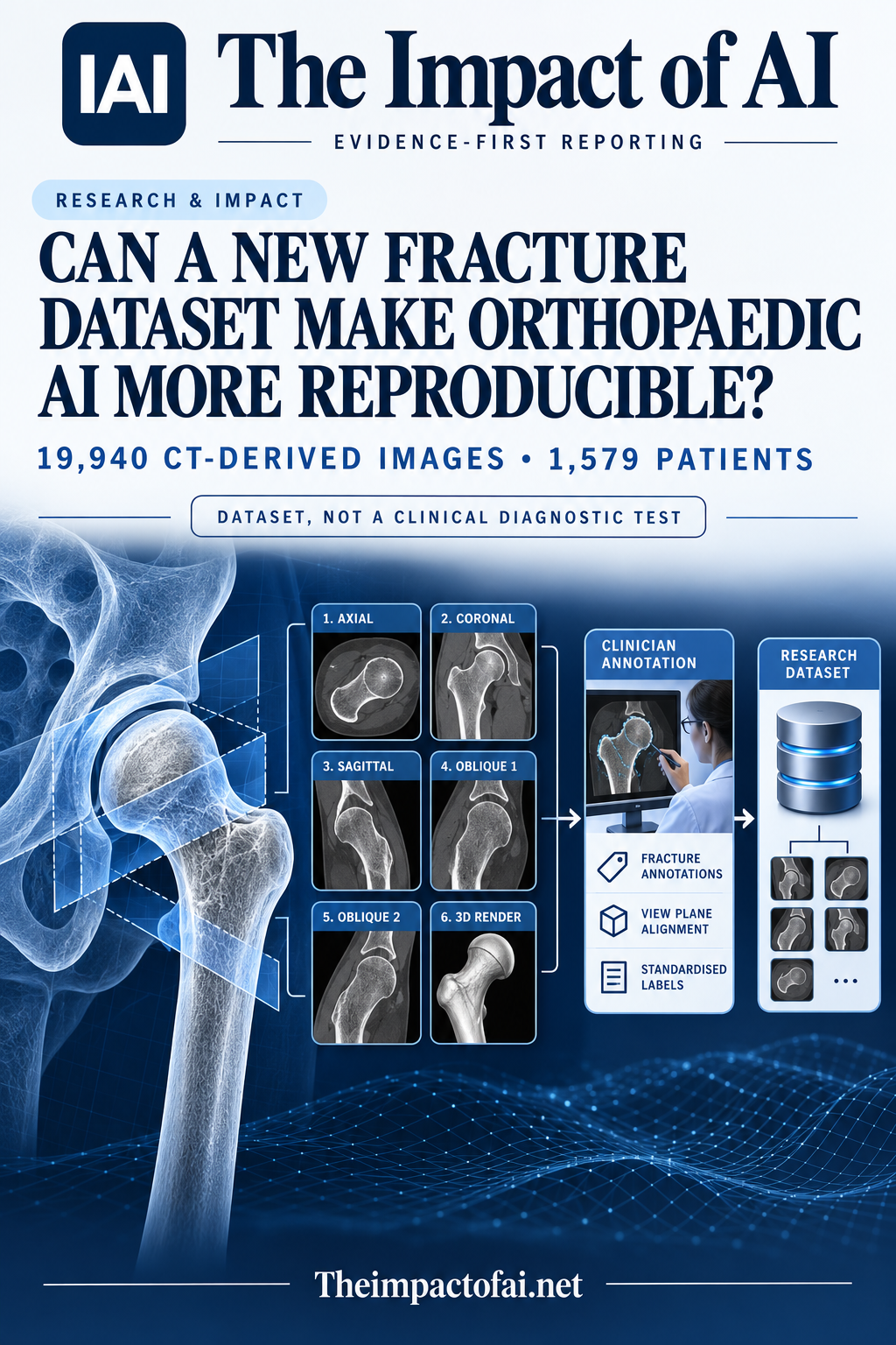
A new open dataset contains 19,940 CT-derived images from 1,579 patients, with multi-view expert labels and patient-level splits. It can support reproducible femoral-neck-fracture research, but its benchmark results are not evidence that an AI system is ready to diagnose or choose treatment.
Editorial responsibility: The Impact of AI Editorial Desk · Report a factual concern
At a glance
- 1The release contains 19,940 CT-derived images from 1,579 patients across axial, coronal and three-dimensional views, each supplied in original and internal-fixator-occlusion-removed forms.
- 2Two senior orthopaedic surgeons annotated the material, with disagreements resolved by a chief surgeon; reported inter-annotator agreement was Cohen’s kappa 0.82 (95% CI 0.78–0.86).
- 3Benchmark AUCs above 0.96 show the selected data can support classification experiments, not that a model is externally validated or clinically safe.
Research topic
A public multi-view CT-derived dataset for development and benchmarking of Garden classification models for femoral neck fractures
The direct answer: a stronger research foundation, not a diagnostic product
The dataset could make orthopaedic-AI experiments easier to reproduce because it supplies a sizeable, publicly available collection with expert labels, multiple views and patient-level partitions. Meiyan Wu and colleagues assembled 19,940 CT-derived images from 1,579 patients with femoral-neck fractures. The release covers axial CT, coronal CT and three-dimensional volume-rendered views, with each view represented both in its original form and in a processed version intended to remove occlusion from internal fixation hardware.
That contribution is infrastructure rather than a clinical result. The article does not show that software improves diagnosis, selects the correct operation, prevents complications or produces better recovery. A model trained on this material would still need testing on untouched data from other hospitals and scanners, followed by prospective evaluation in a real workflow. The high benchmark scores reported in the paper therefore answer whether the dataset contains learnable class information, not whether an autonomous fracture classifier should be used with patients.[1]
What was collected and why the six-view structure matters
The source examinations were retrospective spiral CT studies gathered under ethics approval. From those studies, the team organised six image categories: original and occlusion-removed axial views, original and occlusion-removed coronal views, and original and occlusion-removed three-dimensional renders. Multi-view organisation matters because fracture displacement and alignment can look different across planes, while fixation hardware can obscure the anatomy that a classifier is meant to examine.
Providing both original and processed forms also lets researchers ask a useful methodological question: does removing hardware obstruction improve classification, or does processing introduce information that will not be available reliably in practice? The authors report a segmentation Dice score of 0.962 for the occlusion region and expert confirmation that the processed anatomy remained plausible. Those checks are valuable, but plausibility is not the same as proving that every reconstructed contour preserves clinically decisive detail.[1]
The annotation process reduces one common source of hidden uncertainty
Two senior orthopaedic surgeons labelled the cases independently, and a chief orthopaedic surgeon resolved discrepancies. Inter-annotator agreement reached a Cohen’s kappa of 0.82, with a 95% confidence interval from 0.78 to 0.86. Reporting agreement is important because the Garden system is a human classification framework: an apparently precise machine label can conceal disagreement or ambiguity in the reference standard used for training.
The workflow is more transparent than relying on a single unreviewed label, but adjudication does not eliminate subjectivity. The final label reflects the participating experts and the available views, and other clinicians may still interpret borderline displacement differently. Researchers using the dataset should report performance by Garden class, inspect confusion between adjacent categories and avoid treating the adjudicated label as an infallible biological truth.[1]
Patient-level splitting is a meaningful defence against leakage
The authors partitioned data at patient level. That design prevents images derived from the same person’s examination from appearing in both training and evaluation sets, a serious leakage risk when one CT study produces many related slices and renderings. Without patient-level separation, a model can appear to generalise while recognising repeated anatomy, acquisition artefacts or processing patterns from the same case.
Balanced cohort characteristics across the reported splits provide another useful check, but they do not create an external test. All splits still originate from the same data-building process. Scanner protocols, referral patterns, fracture prevalence, fixation practice and documentation may differ elsewhere. The decisive test will be whether a locked model trained on this release retains calibration and class-specific sensitivity at independent hospitals without tuning on their evaluation labels.[1]
Why AUC above 0.96 should not be read as clinical accuracy
Representative convolutional and transformer architectures achieved areas under the receiver operating characteristic curve above 0.96 on the coronal-view, occlusion-removed subset. This demonstrates strong separation within the benchmark. It does not disclose a single safe operating threshold, nor does an AUC state how many clinically important fractures would be misclassified in each Garden category.
The benchmark used a selected processed subset, whereas clinical deployment could involve inconsistent views, motion, unusual anatomy, different fixation materials and incomplete studies. A high AUC can coexist with poor calibration or concentrated errors in the cases where treatment decisions are most contested. Future evaluations should publish per-class sensitivity, specificity, predictive values at realistic prevalence, calibration and reader studies comparing clinicians with and without model assistance.[1]
What changes for patients—and what evidence is still needed
For patients, the near-term benefit is indirect: open, standardised data can help researchers compare methods on the same cases and inspect failure modes instead of presenting results from inaccessible private collections. The dataset is available through Zenodo under a CC BY 4.0 licence, which supports independent replication and secondary analysis. The work was supported by public research programmes in Chongqing, and the authors declared no competing interests.
Clinical impact would require substantially more evidence. Researchers should validate models on independent hospitals and raw clinical inputs, test whether occlusion removal changes diagnostically relevant structures, examine performance across age, sex, fracture subtype and scanner conditions, and measure agreement with treatment-relevant expert decisions. Prospective studies must then establish whether assistance improves consistency or speed without anchoring clinicians to confident but incorrect classifications. Until then, this is a useful research resource, not a certified diagnostic pathway.[1]
What this means for people
- Open data may reduce duplicated effort and make orthopaedic imaging research easier to audit.
- Models trained on processed views could fail when real clinical images differ from the benchmark.
- Human review remains essential because fracture classification can influence treatment planning.
Global context
The dataset was assembled by institutions in Chongqing, China, and is openly licensed for international research. Its availability improves representation of Chinese clinical imaging in a field often dominated by closed hospital datasets, but openness does not guarantee transportability. Independent teams should test it against local scanners, trauma patterns, reporting practices and treatment pathways before drawing clinical conclusions.
What the evidence does not yet show
- The release is a retrospective dataset and benchmark, not a prospective diagnostic study.
- All splits arise from one data-building process; no independent hospital evaluation is reported.
- Occlusion-removed images are processed reconstructions whose clinical fidelity needs further testing.
- AUC above 0.96 on a selected subset does not reveal a safe threshold or treatment effect.
- Garden labels remain a human-adjudicated reference standard with residual subjectivity.
What to watch next
- Independent validation using raw studies from different hospitals, scanners and populations.
- Per-class errors, calibration and clinically prespecified operating thresholds.
- Reader studies comparing clinicians alone with clinicians assisted by a locked model.
- Prospective evidence on diagnostic consistency, workflow time and downstream treatment decisions.
Living evidence record
Impact record IAI-03IJLJU
Evidence stage
Studied
Confidence
Supported
Reporting basis
Source analysis
Independent or research support
Present
Record status
Monitoring
Last checked
8 October 2026
Source trail
1 direct source across 1 source type.
People impact
Documented in this record.
Uncertainty
Limits and next checks are explicit.
Stages describe the evidence available—not whether a technology is good or bad. See the public method.
Single-source reporting disclosure
This record analyses one direct source. It can establish what Scientific Data published or reported, but it is not independent corroboration of every performance claim or predicted outcome. The confidence label will change only when broader evidence is added.
Evidence trail
Sources used for this report
Links checked 8 October 2026
This report is labelled source analysis. We summarise and analyse source material in our own words; company statements remain attributed claims until independently supported. Translated summaries preserve the meaning of the original source and link back to it. Read our editorial standards.
Continue the story
Related reporting
Health & Life Sciences
Can AI spot liposarcoma on ultrasound?
A peer-reviewed Chinese study reports strong results in a 95-patient internal test, including 0.97 accuracy. But all 317 patients came from one hospital and there was no external validation, so this is a proof of concept—not a clinically cleared diagnostic system.
9 min · 1 source
Health & Life Sciences
Can AI discover new MRI signs for glioblastoma?
It generated eight candidate visual signs from 106 glioblastoma scans, but only one was externally tested. Two radiologists achieved moderate discrimination between 50 glioblastomas and 50 metastases, so this is a discovery signal—not a clinical diagnostic.
7 min · 1 source
Health & Life Sciences
Does 99% MRI accuracy mean an Alzheimer’s diagnostic is ready?
No. A peer-reviewed model classified images from one 6,400-image Kaggle collection with reported 99% test accuracy, but it has no external clinical validation and the published test counts do not reconcile cleanly.
8 min · 2 sources
The Impact Brief
Keep the evidence trail, not the noise.
Get the most consequential AI developments with direct sources and clear limits.
Reader commentary
Add evidence, experience or a question
No account is required. Reader notes are published after a brief civility, relevance and safety check; disagreement is welcome.
Explore commentary across the portal →Published reader notes
0No published reader notes yet. You can start the evidence-led discussion above.
Prefer a private correction or response? Contact the newsroom.