Can this MRI model draw brain-tumour boundaries reliably?
A peer-reviewed multimodal segmentation model was developed on 2,422 public MRI volumes and externally tested on 125 cases. Accuracy remained useful but fell outside the development data; no prospective clinical workflow, radiologist comparison or patient-outcome test was performed.
Editorial responsibility: The Impact of AI Editorial Desk · Report a factual concern
Research topic
Whether modality-weighted fusion and attention improve automated segmentation of brain-tumour regions across internal and external MRI datasets
At a glance
- 1After excluding incomplete or failed-quality cases, the team used 2,422 public MRI volumes for development: 1,600 for training, 400 for validation and 422 for internal testing, with partitions made at patient level and stratified by source.
- 2The full model achieved a mean Dice score of 0.815 internally across whole tumour, tumour core and enhancing tumour, then 0.782 on an external 125-case BraTS 2021 cohort—a 0.033 absolute drop under domain shift.
- 3The study did not test live radiology or treatment workflows, compare assistance against clinicians, measure patient outcomes, or establish reliability when MRI sequences are missing.
The Impact Brief
Keep the evidence trail, not the noise.
Get the most consequential AI developments with direct sources and clear limits.
Living evidence record
Impact record IAI-1PL1853
Evidence stage
Studied
Confidence
Supported
Reporting basis
Source analysis
Independent support
Present
Record status
Monitoring
Last checked
3 October 2026
Source trail
2 direct sources across 2 source types.
People impact
Documented in this record.
Uncertainty
Limits and next checks are explicit.
Stages describe the evidence available—not whether a technology is good or bad. See the public method.
Related-source reporting disclosure
This record analyses 2 linked source records around the same underlying development. The extra records add method, date or context, but they do not by themselves constitute independent replication of every performance claim or predicted outcome.
Four MRI streams were weighted before segmentation
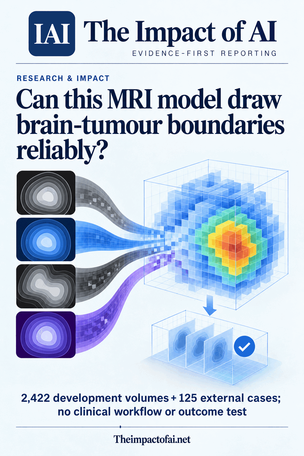
Brain-tumour segmentation asks a model to label every voxel belonging to clinically relevant tumour regions. The new AMF-U-Net study combines four standard MRI sequences—T1, contrast-enhanced T1, T2 and FLAIR—because each reveals different tissue characteristics. A modality-fusion module learns a softmax weight for each stream before features enter a three-dimensional residual U-Net.
Attention gates then suppress less relevant encoder features as the network reconstructs the segmentation. The training objective combines Dice loss, which rewards overlap, with categorical cross-entropy, which penalises class errors. The output divides tumour into whole tumour, tumour core and enhancing tumour regions rather than making a simple tumour-or-no-tumour classification.
This architecture is a technical proposal, not a diagnosis. Segmentation could eventually support measurement, surgical planning or treatment monitoring, but the paper evaluates overlap with dataset labels. It does not show that the boundaries alter a radiologist’s report, improve a treatment decision or lead to better survival, function or quality of life.[1]
The development set contained 2,422 usable public volumes
The authors began with 1,846 BraTS 2023 volumes and 650 UCSF-PDGM volumes. They excluded 28 BraTS cases and 19 UCSF cases with missing modalities, then removed 18 and nine respectively after quality control. That left 1,800 BraTS and 622 UCSF-PDGM volumes, or 2,422 in total.
Partitions were made at patient level and stratified by data source. Training used 1,600 volumes, validation 400 and the internal test 422. The paper further describes balanced development groups across glioma, meningioma, paediatric and secondary-tumour cohorts, with 400 training cases from each. Patient-level partitioning reduces direct leakage between train and test sets, while source stratification reduces the chance that one source dominates a split.
However, every usable development case had all four modalities. Real-world imaging can be incomplete, corrupted or acquired with different protocols. Excluding incomplete cases creates a cleaner task and means the reported performance does not establish resilience when a sequence is missing—the very condition that could determine whether a multimodal system works during routine care.[1][2]
Internal overlap was strongest for the whole tumour
On the 422-case internal test, Dice overlap was 0.845 for whole tumour, 0.813 for tumour core and 0.788 for enhancing tumour, producing a three-region mean of 0.815. Dice ranges from zero for no overlap to one for perfect overlap. The lower enhancing-tumour score shows that the smaller, clinically important region remained harder to delineate.
The paper compares the model with 3D U-Net, nnU-Net, UNETR and Swin UNETR trained on the same split. It also runs ablations over fusion and attention components. Against simple concatenation, the modality-fusion module improved mean Dice by 0.0336, with a Holm-adjusted p value of 1.44×10⁻⁷. Against a cross-attention fusion variant, the gain was 0.0073, adjusted p=0.0180.
Same-split comparisons are more informative than borrowing headline scores from unrelated papers, but extensive architectural choices and ablations can still tune a research programme to one benchmark. Statistical significance across cases does not automatically mean a boundary change is clinically important. The paper reports overlap and distance metrics, not whether specialists would accept or correct each contour.[1]
External testing exposed a measurable domain shift
The authors then tested 125 BraTS 2021 cases outside the development partition. Dice was 0.823 for whole tumour, 0.784 for tumour core and 0.739 for enhancing tumour, with a mean of 0.782. The mean fell by 0.033 from the internal result, and the enhancing region again performed worst.
That external cohort is one of the paper’s most valuable features because it tests transfer beyond the immediate training and validation data. Yet 125 curated challenge cases are still not the same as prospective, consecutive patients across hospitals. Dataset conventions, expert labels and preprocessing can remain related even when the release year changes.
A stronger clinical validation would freeze the model, recruit multiple independent centres, retain difficult and incomplete scans, pre-specify failure criteria and report results by tumour type, size, scanner, institution and demographic group. It should compare the model with radiologists and measure how often assistance saves time, changes a decision or creates a harmful correction burden.[1]
The paper documents compute and deployment limits
Training used a high-end NVIDIA RTX 4090 graphics processor, and the authors acknowledge computational cost. Three-dimensional multimodal models can demand memory, storage and preprocessing capacity that are not evenly available across hospitals. Inference time, energy use and integration effort were not evaluated as deployment outcomes.
The study also did not report calibration of voxel-level uncertainty, a prospective failure-detection mechanism or robust handling of missing modalities. A contour can look plausible while being wrong at a clinically important margin. Systems intended for supervised use need a way to surface low confidence and preserve an auditable route back to the underlying images and human correction.
The public-dataset design meant no new participants were enrolled and the paper says human-subject approval was not applicable. That supports reproducibility and avoids exposing new patient information, but it does not test consent, workflow, accountability or cybersecurity questions that arise when a model is connected to hospital systems.[1]
Disclosures contain two points that merit clarification
The authors are affiliated with institutions in Saudi Arabia, India and South Korea and declare no competing interests. The acknowledgements say the work was supported by South Korea’s National IT Industry Promotion Agency through a grant for a physical-AI factory model, while the separate funding statement says no funding was received. Readers need a corrected version to reconcile those declarations.
The data-availability section also describes UCSF-PDGM with language associated with brain metastases, even though the methods and official Cancer Imaging Archive record identify it as a preoperative diffuse-glioma dataset. That appears to be an editorial inconsistency rather than a change to the reported sample, but dataset identity is central to judging generalisability and should be clarified in the final version.
The journal labels the article a peer-reviewed accepted version published on 3 October 2026 and notes that further copy-editing and typesetting will precede the version of record. The funding and dataset wording should be checked again when that final article appears. Neither issue by itself overturns the segmentation results, but both affect confidence in documentation.[1][2]
The next test is whether assistance improves real care
For radiologists and oncology teams, the model is best understood as a research candidate for supervised contouring. The external result suggests useful transfer while also demonstrating degradation. Any pilot should preserve manual review, record edits and failures, and keep clinical responsibility with qualified staff rather than treating the output as an autonomous boundary.
For patients, overlap scores do not answer the questions that matter most: whether a tumour is missed, whether an operation or radiotherapy plan changes safely, whether repeat scanning is avoided and whether outcomes improve. Performance also needs to be tested across age, sex, geography, tumour rarity and scanner access so that error is not concentrated in underrepresented groups.
Confidence would rise with preregistered, prospective multicentre evaluation against specialist readers; tests with missing and lower-quality sequences; calibrated uncertainty; and workflow outcomes such as contouring time, correction burden, inter-reader agreement and treatment-plan changes. Until then, AMF-U-Net advances a benchmarked segmentation method, not a proven clinical service.[1]
What this means for people
- Automated contours could reduce repetitive specialist work, but inaccurate margins may add correction burden or influence high-stakes treatment planning.
- Patients need evidence about missed tumours, treatment decisions and outcomes—not only average overlap on public datasets.
- Compute, scanner and data differences may limit transfer to hospitals outside well-resourced research settings unless prospective testing includes them.
Global context
The research collaboration spans Saudi Arabia, India and South Korea and relies on public North American and international challenge datasets. That is broader than a single-institution study, but it does not establish performance in Middle Eastern, South Asian or other local clinical populations. Prospective evaluation should include hospitals with different scanners, protocols, disease mixes and computing resources rather than assuming a public benchmark represents global practice.
What the evidence does not yet show
- The external test used 125 curated BraTS 2021 cases rather than prospective consecutive patients from independent clinical workflows.
- Cases missing any of four MRI modalities were excluded, so robustness to incomplete routine imaging remains unproven.
- The study measured segmentation overlap and distance, not radiologist performance, treatment decisions, patient outcomes or adverse events.
- Architecture selection and ablations occurred within the same research programme; wider independent replication is needed.
- The accepted article contains an apparent funding-statement inconsistency and imprecise wording about the UCSF-PDGM dataset that should be corrected in the final version.
What to watch next
- Prospective multicentre tests using consecutive patients, varied scanners and frozen preprocessing and model weights.
- Reader studies measuring correction burden, reporting time, missed regions and effects on surgery or radiotherapy planning.
- Robustness to missing modalities, low-quality acquisitions and rare tumour presentations, with calibrated uncertainty and failure alerts.
- Clarification of the funding acknowledgement and UCSF-PDGM description in the final version of record.
Evidence trail
Sources used for this report
Links checked 3 October 2026
This report is labelled source analysis. We summarise and analyse source material in our own words; company statements remain attributed claims until independently supported. Translated summaries preserve the meaning of the original source and link back to it. Read our editorial standards.
Continue the story
Related reporting
Health & Life Sciences
Can AI support breast-ultrasound decisions across countries?
A peer-reviewed South Korean-led study validated an interpretable retrieval-augmented system across 8,311 images from 11 cohorts in seven countries and tested assistance with four readers. The retrospective evidence is encouraging, but it is not a prospective screening trial or proof of better patient outcomes.
8 min · 3 sources
Health & Life Sciences
Can brain MRI reveal more than BMI?
A peer-reviewed study trained a deep-learning model on 45,702 MRI scans from six cohorts. Brain-derived features tracked BMI and separated several disease groups better than BMI alone—but the disease analysis stayed inside UK Biobank and cannot establish cause, diagnosis or clinical benefit.
9 min · 2 sources
Health & Life Sciences
Can clinical AI recognise when the patient record does not support an answer?
A new clinical-agent benchmark raises a practical question for health systems: can an assistant explain what the record cannot establish? Our analysis examines evidence, local testing and the burden on staff.
6 min · 2 sources
Reader discussion
Add evidence, experience or a question
No account is required. Reader notes are published after a brief civility, relevance and safety check; disagreement is welcome.
Published reader notes
0No published reader notes yet. You can start the evidence-led discussion above.
Prefer a private correction or response? Contact the newsroom.