A lung-nodule model travelled better—but its labels were not pathology
A Chinese research team improved discrimination and calibration when moving a CT model from a US dataset to a Portuguese cohort. The test covered 618 external nodules, but remained retrospective and partly relied on radiologist ratings.
The short version
3 key points
- What changed
- GWDRO-Lung was developed on 1,842 nodules from 1,003 patients in the US LIDC-IDRI dataset and tested externally on 618 nodules from 284 patients in Portugal's LNDb dataset.
- Why it matters
- A better calibrated score could reduce avoidable invasive work-ups without missing more cancers, but that benefit was only modelled here. Patients still need radiologist judgement, follow-up context and pathology where clinically indicated.
- What to watch
- Prospective multicentre pathology-confirmed validation, stable prespecified thresholds, public code, calibration-drift monitoring, single-case deployment tests and comparisons that add clinical and molecular information.
The model was built for the point where medical AI often fails
A lung-nodule score can look strong where it was trained and then become less accurate or overconfident when scanners, protocols or patient populations change. Researchers in China designed GWDRO-Lung to align the internal structure of different CT cohorts while training a risk score to remain calibrated. Development used 1,842 nodules from 1,003 patients in the US LIDC-IDRI collection, divided into three acquisition-defined cohorts. External testing used 618 nodules from 284 patients in Portugal's independent LNDb collection.[1]
Why calibration matters to a person facing a biopsy
Clinicians act on risk thresholds: a score may lead to surveillance, more imaging or an invasive work-up. The paper's retrospective decision-curve analysis reported higher net benefit across 10%–40% thresholds. At 20%, the model was estimated to avoid roughly eight additional unnecessary biopsies per 100 nodules at the same weighted trade-off. That is a modelled comparison on existing data; no patient actually had care changed by the system.[1]
Who it affects
A better calibrated score could reduce avoidable invasive work-ups without missing more cancers, but that benefit was only modelled here. Patients still need radiologist judgement, follow-up context and pathology where clinically indicated.[1]
Keep in mind
Retrospective public datasets; radiologist-rated rather than pathology-confirmed LIDC labels; one external source; synthetic protocol shift; image-only inputs; five training seeds; no prospective care or patient-outcome test.
Source analysis · 1 linked source. Original source date: 17 October 2026.
Continue the story
Read next
Health & Life Sciences
An Alzheimer’s MRI model scored 94.5%. On new data, it fell to 61.6%
For a patient, the important test is whether a model works beyond familiar images. This classifier’s accuracy dropped sharply on an external dataset, and it remains a research system rather than a clinical diagnostic tool.
2 min · 1 source
Health & Life Sciences
Can brain MRI reveal more than BMI?
A peer-reviewed study trained a deep-learning model on 45,702 MRI scans from six cohorts. Brain-derived features tracked BMI and separated several disease groups better than BMI alone—but the disease analysis stayed inside UK Biobank and cannot establish cause, diagnosis or clinical benefit.
2 min · 2 sources
Read more The full report, methods and context
The model was built for the point where medical AI often fails
A lung-nodule score can look strong where it was trained and then become less accurate or overconfident when scanners, protocols or patient populations change. Researchers in China designed GWDRO-Lung to align the internal structure of different CT cohorts while training a risk score to remain calibrated. Development used 1,842 nodules from 1,003 patients in the US LIDC-IDRI collection, divided into three acquisition-defined cohorts. External testing used 618 nodules from 284 patients in Portugal's independent LNDb collection.
Across five matched training seeds, GWDRO-Lung achieved external AUROC of 0.878, compared with 0.857 for the strongest recent imaging baseline. Expected calibration error—how far predicted probabilities depart from observed frequencies—was 0.066 versus 0.106. Worst-held-out-cohort AUROC was 0.851 versus 0.829. These are modest but consistent improvements in ranking and probability quality, not a claim of perfect classification.[1]
Why calibration matters to a person facing a biopsy
Clinicians act on risk thresholds: a score may lead to surveillance, more imaging or an invasive work-up. The paper's retrospective decision-curve analysis reported higher net benefit across 10%–40% thresholds. At 20%, the model was estimated to avoid roughly eight additional unnecessary biopsies per 100 nodules at the same weighted trade-off. That is a modelled comparison on existing data; no patient actually had care changed by the system.
The authors also created a controlled stress set by resampling 612 held-out scans to thicker slices and sharper reconstruction kernels. Performance held up better than comparison models, and ablations linked the largest external-AUROC loss to removal of the cohort-alignment component. Yet the strongest configuration benefits from seeing several target nodules together, adding a practical dependency that a single-case clinical workflow may not satisfy.[1]
The distinction that matters
From development scans to an external CT cohort
The paper tested whether the model preserved both ranking and probability reliability after a dataset change.
Develop
1,842 US nodules across three cohorts
Learn CT features, cohort structure and calibration
Transfer
Test 618 Portuguese nodules
AUROC 0.878; calibration error 0.066
Limit
Retrospective, imperfect reference labels
Prospective pathology-confirmed validation still required
The reference standard is the largest unresolved weakness
LIDC-IDRI labels were derived from radiologists' malignancy ratings, not pathology-confirmed outcomes. The paper's own noise analysis found that the advantage narrowed as labels became less reliable. External LNDb data came from one source, and the protocol shift was synthetic. The study was image-only, retrospective and did not test prospective referral, biopsy or survival outcomes. Its article-in-press text said code would be released after acceptance, but did not yet link that release.
For radiologists and patients, the result is therefore a promising engineering step rather than a deployable cancer test. The assessment would change with prospective multicentre evaluation against pathology-confirmed outcomes, prespecified thresholds, full calibration monitoring and comparisons that include clinical variables. Until then, the evidence supports further validation of the transport-and-calibration method—not autonomous nodule triage.[1]
Watch the background, then check the evidence
Illustrative photo · not a video frame
The Royal Society · 44 min 28 sec · Background explainer
Is this the AI future we want? | The Royal Society
YouTube loads only when you choose; playback starts when you press play.
A Royal Society discussion with Madhumita Murgia and biomedical engineer Alison Noble on AI’s potential in healthcare, science and culture, recorded at the 30 June 2026 event.
Evidence check: A discussion of opportunities and risks, including future possibilities. It is not evidence that every application described is available or clinically proven.
Published 2026-07-01. Source checked 14 October 2026. Check captions on the publisher’s video page.
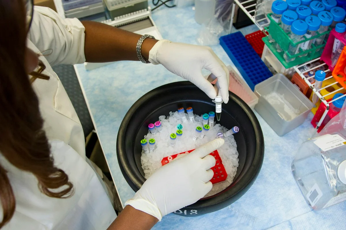
Check the healthcare evidencePhoto: Daniel Sone / National Cancer Institute / Unsplash · Licence
Evidence check
Retrospective public datasets; radiologist-rated rather than pathology-confirmed LIDC labels; one external source; synthetic protocol shift; image-only inputs; five training seeds; no prospective care or patient-outcome test.
Reporting basis: Source analysis. Original source date: 17 October 2026.
Explore the full source and evidence record
What this means for people
- A better calibrated score could reduce avoidable invasive work-ups without missing more cancers, but that benefit was only modelled here. Patients still need radiologist judgement, follow-up context and pathology where clinically indicated.
Global context
The authors are based in China, development images came from a US consortium and external images came from Portugal. That cross-country design is useful, but one external dataset cannot represent the scanner, population and workflow variation across screening programmes worldwide.
What the evidence does not yet show
- Retrospective public datasets; radiologist-rated rather than pathology-confirmed LIDC labels; one external source; synthetic protocol shift; image-only inputs; five training seeds; no prospective care or patient-outcome test.
What to watch next
- Prospective multicentre pathology-confirmed validation, stable prespecified thresholds, public code, calibration-drift monitoring, single-case deployment tests and comparisons that add clinical and molecular information.
Living evidence record
Impact record IAI-1KGI22Q
Evidence stage
Studied
Confidence
Supported
Reporting basis
Source analysis
Independent or research support
Present
Record status
Monitoring
Last checked
18 October 2026
Source trail
1 direct source across 1 source type.
People impact
Documented in this record.
Uncertainty
Limits and next checks are explicit.
Stages describe the evidence available—not whether a technology is good or bad. See the public method.
Single-source reporting disclosure
This record analyses one direct source. It can establish what npj Digital Medicine published or reported, but it is not independent corroboration of every performance claim or predicted outcome. The confidence label will change only when broader evidence is added.
Evidence trail
Sources used for this report
Links checked 18 October 2026
Research question, topics and regions
Whether relation-level cohort alignment improves lung-nodule malignancy ranking and probability calibration after CT dataset shift
Topics: Lung cancer · Medical imaging · External validation · Calibration · Dataset shift
Regions: China · United States · Portugal · Asia · Europe · North America · Global
This report is labelled source analysis. We summarise and analyse source material in our own words; company statements remain attributed claims until independently supported. Translated summaries preserve the meaning of the original source and link back to it. Read our editorial standards.
The Impact Brief · Free
Keep following the stories that matter to you.
Get our weekday briefing on what changed and why it matters. Choose your topics below.
Reader commentary
What does this mean where you are?
Tell us what you have seen at work, in your community or in everyday life. A question is welcome too. No account is needed; we review notes before publishing them.
Explore commentary across the portal →Published reader notes
0No published reader notes yet. You can start the evidence-led discussion above.
Prefer a private correction or response? Contact the newsroom.