A pathology AI ensemble recognised Barrett’s cancer—but has not yet improved care
A peer-reviewed benchmark combined five pathology foundation models across magnifications. It performed strongly on 3,528 archived slides, but remains a retrospective classifier rather than a tested clinical diagnostic.
The short version
- What happened
- The benchmark used 3,528 digitised tissue slides from 790 patients, covering tissue from normal appearance through Barrett's changes to oesophageal adenocarcinoma.
- Who it affects
- For people undergoing Barrett's surveillance, a reliable second-read tool could eventually help pathology teams prioritise suspicious samples. This paper does not show that it reduces missed cancer, unnecessary treatment or waiting time in routine care.
- What remains unproven
- The study was retrospective and slide based; it did not prospectively evaluate pathologists working with the tool.
Combining views helped more than trusting one model
The team tested pathology foundation models on haematoxylin-and-eosin slides viewed at magnifications from 5× to 40×. It then combined the five single-scale models whose predictions agreed most closely with experts. The aim was to capture both broad tissue structure and cellular detail rather than rely on one representation.[1][2]
Accuracy did not mean every category was equally reliable
The denominator matters. The dataset contained 3,528 whole-slide images from 790 patients and represented stages from normal tissue through metaplasia and dysplasia to cancer. Overall accuracy can look high when some categories are more common or easier, which is why the lower macro F1 score for morphological subtyping deserves equal attention.[2]
Who it affects
For people undergoing Barrett's surveillance, a reliable second-read tool could eventually help pathology teams prioritise suspicious samples. This paper does not show that it reduces missed cancer, unnecessary treatment or waiting time in routine care.[1][2]
Keep in mind
The study was retrospective and slide based; it did not prospectively evaluate pathologists working with the tool.
Reported accuracy and AUROC are not patient outcomes, and overall accuracy can obscure weaker performance in rarer morphology classes.
The material used a particular fixation and digitisation workflow, so external sites need validation across their own laboratories and scanners.
No effect on diagnostic decisions, treatment, workload, safety or survival was measured.
Source analysis · 2 linked sources. Original source date: 11 November 2025. Latest supporting source: 21 October 2026.
Keep following the evidence
Join The Impact Brief founding list for selected AI reports, their effect on people and the evidence limits. Read a sample first.
Read the full report · 3 min The evidence, methods, implications and limitations

Combining views helped more than trusting one model
The team tested pathology foundation models on haematoxylin-and-eosin slides viewed at magnifications from 5× to 40×. It then combined the five single-scale models whose predictions agreed most closely with experts. The aim was to capture both broad tissue structure and cellular detail rather than rely on one representation.
For cancer detection, the ensemble reported an area under the receiver-operating curve of 0.909, an F1 score of 0.836 and accuracy of 0.959. The morphology task was harder: its corresponding figures were 0.907, 0.696 and 0.795, with kappa agreement of 0.651.[1][2]
Accuracy did not mean every category was equally reliable
The denominator matters. The dataset contained 3,528 whole-slide images from 790 patients and represented stages from normal tissue through metaplasia and dysplasia to cancer. Overall accuracy can look high when some categories are more common or easier, which is why the lower macro F1 score for morphological subtyping deserves equal attention.
Agreement with pathologists is useful for prioritising suspicious slides, but it is not the same as proving a safer diagnosis. The study did not randomise clinical readers, compare final treatment decisions or test whether a missed or overcalled category changed what happened to a patient.[2]
The distinction that matters
From one tissue slide to an ensemble decision
The system aggregates information across image scales and model backbones before producing a slide-level classification.
Digitise
Turn each tissue slide into image tiles
3,528 slides from 790 patients
Compare
Run models from 5× to 40×
Different tissue and cellular views
Ensemble
Combine five expert-consistent models
One slide-level prediction
A benchmark, not a deployment verdict
The experiment used archived, specially fixed and digitised slides. Performance transferred across fixation conditions without task-specific fine-tuning in the reported tests, but a pathology service would still need external validation on its scanners, staining, specimen mix and workflow before relying on the model.
The authors declared no competing interests in the public preprint. The next persuasive evidence would be a prospective, multi-centre reader study with pre-specified error categories, calibration and patient-relevant outcomes. Until then, the ensemble shows that model diversity can improve a benchmark—not that it can replace expert pathology review.[1][2]
Watch the background, then check the evidence
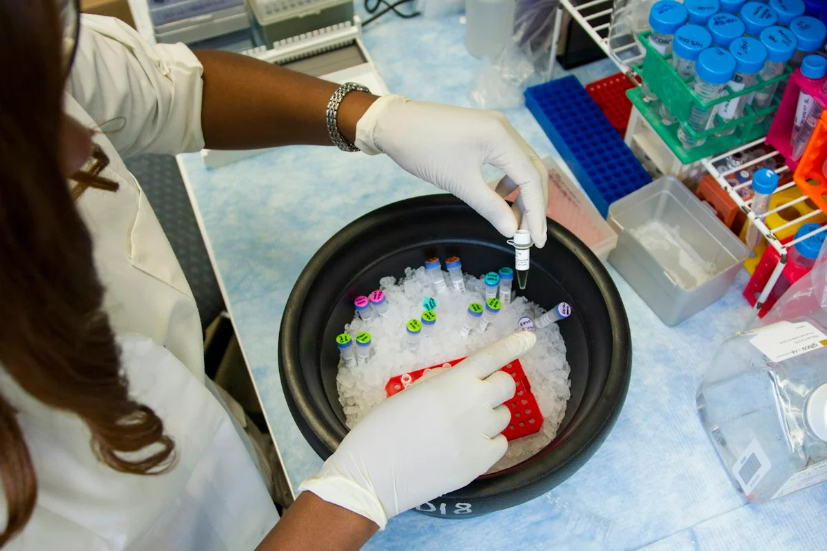
Illustrative photo · not a video frame
The Royal Society · 44 min 28 sec · Background explainer
Is this the AI future we want? | The Royal Society
YouTube loads only when you choose; playback starts when you press play.
A Royal Society discussion with Madhumita Murgia and biomedical engineer Alison Noble on AI’s potential in healthcare, science and culture, recorded at the 30 June 2026 event.
Evidence check: A discussion of opportunities and risks, including future possibilities. It is not evidence that every application described is available or clinically proven.
Published 2026-07-01. Source checked 14 October 2026. Check captions on the publisher’s video page.
Check the healthcare evidencePhoto: Daniel Sone / National Cancer Institute / Unsplash · Licence
Evidence check
The study was retrospective and slide based; it did not prospectively evaluate pathologists working with the tool.
Reported accuracy and AUROC are not patient outcomes, and overall accuracy can obscure weaker performance in rarer morphology classes.
The material used a particular fixation and digitisation workflow, so external sites need validation across their own laboratories and scanners.
No effect on diagnostic decisions, treatment, workload, safety or survival was measured.
Reporting basis: Source analysis. Original source date: 11 November 2025. Latest supporting source: 21 October 2026.
Continue the story
Read next
Continue exploring digital pathology: compare the finding and its evidence limits.
Can AI predict five-year colorectal cancer survival from pathology slides?
A retrospective preprint reports moderate discrimination across a US trial cohort and an external NHS dataset. The model is not ready to guide treatment, and its selected ensemble did not clearly beat the strongest comparator.
Read the next reportExplore the full source and evidence record
What this means for people
- For people undergoing Barrett's surveillance, a reliable second-read tool could eventually help pathology teams prioritise suspicious samples. This paper does not show that it reduces missed cancer, unnecessary treatment or waiting time in routine care.
Global context
The work was led by German and Iranian academic researchers and addresses a specialised digital-pathology setting. Laboratories without whole-slide scanners, compatible fixation or local validation would face different operational and equity constraints.
What the evidence does not yet show
- The study was retrospective and slide based; it did not prospectively evaluate pathologists working with the tool.
- Reported accuracy and AUROC are not patient outcomes, and overall accuracy can obscure weaker performance in rarer morphology classes.
- The material used a particular fixation and digitisation workflow, so external sites need validation across their own laboratories and scanners.
- No effect on diagnostic decisions, treatment, workload, safety or survival was measured.
What to watch next
- External validation on independently collected slides from multiple pathology services
- A prospective reader study comparing pathologists with and without the ensemble
- Class-specific false-negative rates, calibration and performance on difficult low-grade dysplasia
- Measured effects on turnaround time, treatment decisions and patient outcomes
Living evidence record
Impact record IAI-0ULRJVA
Evidence stage
Studied
Confidence
Supported
Reporting basis
Source analysis
Independent or research support
Present
Record status
Monitoring
Last checked
21 October 2026
Source trail
2 direct sources across 1 source type.
People impact
Documented in this record.
Uncertainty
Limits and next checks are explicit.
Stages describe the evidence available—not whether a technology is good or bad. See the public method.
Related-source reporting disclosure
This record analyses 2 linked source records around the same underlying development. The extra records add method, date or context, but they do not by themselves constitute independent replication of every performance claim or predicted outcome.
Evidence trail
Sources used for this report
Links checked 21 October 2026
Research question, topics and regions
Whether an ensemble of pathology foundation models can classify Barrett's oesophagus morphology and detect oesophageal adenocarcinoma on archived whole-slide images
Topics: Digital pathology · Cancer detection · Barrett's oesophagus · Foundation models · Clinical validation
Regions: Germany · Iran
This report is labelled source analysis. We summarise and analyse source material in our own words; company statements remain attributed claims until independently supported. Translated summaries preserve the meaning of the original source and link back to it. Read our editorial standards.
Reader commentary
What does this mean where you are?
Tell us what you have seen at work, in your community or in everyday life. A question is welcome too. No account is needed; we review notes before publishing them.
Explore commentary across the portal →Published reader notes
0No published reader notes yet. You can start the evidence-led discussion above.
Prefer a private correction or response? Contact the newsroom.