Can routine implant checks detect a cochlear electrode moving out of place?
A four-centre study detected 14 of 16 confirmed migrations from routine electrical telemetry and produced no false alarms in 16 matched controls. The 32-case proof-of-concept used one manufacturer's implant family and needs prospective, independent validation.
The short version
- What happened
- The retrospective evaluation matched 16 radiologically confirmed migration cases with 16 stable controls drawn from four cochlear-implant centres; patients ranged from 1 to 89 years old.
- Who it affects
- Children and adults could eventually receive an additional low-burden check from telemetry already collected during implant programming, but the method is not ready to replace imaging.
- What remains unproven
- Only 32 evaluated implant cases: 16 confirmed migrations and 16 matched stable controls
Keep in mind
Only 32 evaluated implant cases: 16 confirmed migrations and 16 matched stable controls
Case-control sampling with 50% migration prevalence cannot provide routine-clinic predictive values
Retrospective and temporally asynchronous telemetry and imaging; no prospective alert timing
MED-EL FLEX-series arrays only, with a training set dominated by one array type
No hearing, quality-of-life, revision, radiation-reduction or cost outcome
Manufacturer advisory roles and external grants disclosed by several authors
Source analysis · 1 linked source. Primary analysed source date: 4 November 2026.
Keep following the evidence
Join The Impact Brief founding list for selected AI reports, their effect on people and the evidence limits. Read a sample first.
Read the full report · 9 min The evidence, methods, implications and limitations
The direct answer: the signal was detectable, but this was a 32-case proof-of-concept
Researchers found that changes in routine electrical telemetry could identify most confirmed cases in which a cochlear implant's electrode array had shifted after surgery. Their machine-learning method correctly flagged 14 of 16 migration cases and classified all 16 matched stable controls as stable. That corresponds to 88% sensitivity, 100% specificity and a receiver-operating-characteristic area of 94% in the study set. The result is promising because telemetry is already collected during ordinary implant checks, whereas confirming electrode position usually requires an X-ray, CT or cone-beam CT scan.
The denominator matters. This was not a screening programme applied to an unselected clinic population. Investigators first identified 16 people or implanted ears with radiologically confirmed migration, then matched each case to one stable control by factors including electrode type, age at surgery, sex and implanted side. That design creates a deliberately balanced set in which half the cases have the rare complication. Sensitivity and specificity can be estimated, but the study cannot tell a clinic how many alerts would occur when migration is uncommon. A positive predictive value calculated from this 50% prevalence would not describe routine care.[1]
What was measured and how the algorithm made a decision
Cochlear implants use an electrode array placed inside the cochlea to stimulate the auditory nerve. If the array moves outward, some contacts can leave their intended position and hearing performance can deteriorate. The researchers used transimpedance matrices: electrical measurements produced by stimulating one contact and recording voltage patterns across others. Those patterns contain information about current flow and therefore about where the most basal electrode sits. The model also used cochlear dimensions and implant parameters, and returned an estimated longitudinal position in millimetres for each telemetry recording.
The team treated the first available position estimate as a baseline. A later estimated movement of more than one millimetre triggered a migration flag, after which the new estimate became the baseline for further checks. The threshold was chosen to exceed expected noise in the radiological reference and was roughly half the spacing between contacts on the most common array. It therefore has a practical measurement rationale, but it is still a study-defined threshold rather than a clinically validated intervention point. Transverse movement toward the inner or outer cochlear wall was not assessed.[1]
The distinction that matters
From routine telemetry to a migration alert
The proposed method turns repeated electrical measurements into an estimated change in the basal electrode's longitudinal position.
Baseline
Estimate position from the first usable telemetry recording
Patient-specific reference
Follow-up
Combine transimpedance patterns, anatomy and implant parameters
New estimated position
Threshold
Compare the change with a one-millimetre boundary
Stable or migration flag
Clinical review
Investigate an alert or continuing concern
Imaging and treatment still require clinician judgment
The comparison used radiology as ground truth across four centres
The retrospective cases came from four cochlear-implant centres and involved surgery between 2011 and 2025. Eligibility required at least two radiological scans of the same implanted ear at different postoperative times. Twenty cases had three-dimensional CT or cone-beam CT imaging and 12 had X-ray projections. Investigators compared those scans to establish whether the basal electrode had moved by more than one millimetre. They excluded several abnormal cochlear conditions and discarded telemetry recordings containing short- or open-circuit faults identified by the manufacturer's software.
The final 32 implant cases represented 28 people, including four bilateral implantations. Ages at surgery ranged from one to 89 years, with a median of 56; 13 patients were female and 15 male. The model identified migration on average 10.6 months after surgery among the 14 detected cases, with a range from about 0.2 to 41.7 months. Those figures describe when the model first crossed its threshold in stored telemetry, not a prospective warning delivered to a clinician. Imaging was usually obtained because of clinical need rather than on the same day as telemetry, so temporal precision could not be established.[1]
Four errors in a larger clinic could change the practical value
The confusion matrix was 14 true positives, two false negatives, 16 true negatives and no false positives. Both missed migrations are clinically important: a screening method that does not alert cannot replace clinical judgment or imaging when hearing changes, device measurements or symptoms remain concerning. Conversely, observing no false positives in only 16 stable controls does not prove that false alarms will remain rare. Even a highly specific test can generate many unnecessary scans or appointments when it is applied repeatedly to thousands of people and the target condition is uncommon.
The authors evaluated each implant case as positive if the system detected at least one event at any point. They did not require the alert to occur between the two scans that bounded the actual migration interval, because telemetry and imaging were asynchronous. That makes the analysis suitable for showing that the electrical pattern contains positional information, but not for showing how early or reliably a clinic would be warned. A prospective study should predefine alert timing, count every alert per person, record follow-up imaging and report whether earlier review changes revision surgery, hearing performance or distress.[1]
A separate training set and one implant family limit generalisation
The position-estimation model had been trained on 179 cases, of which 162 used one MED-EL FLEX 28 array. In out-of-fold evaluation, the selected random-forest model estimated basal-electrode position with a mean absolute error of 0.94 millimetres and a standard deviation of 0.75 millimetres; errors ranged from minus 3.5 to plus 2.4 millimetres. Because the migration threshold was one millimetre, prediction error and the decision boundary are close. The longitudinal patterns across repeated measurements appear to carry useful information, but clinicians should not interpret an estimated millimetre value as exact anatomy.
All evaluated devices were lateral-wall FLEX-series arrays from the same manufacturer. The result therefore does not establish performance for pre-curved arrays, other electrode geometries, different telemetry protocols or other manufacturers' software. Centre variation is a strength, but the selected cases had unusually rich imaging follow-up and may not resemble people receiving ordinary care. The data are available only from the corresponding author on reasonable request, which makes independent reanalysis harder than an open benchmark would be.[1]
What the finding could mean for children and long-term follow-up
A radiation-free check could be particularly useful for children, who may have decades of follow-up and for whom minimising unnecessary imaging is valuable. Telemetry is quick and already built into implant programming, so an alert might help audiologists or clinical engineers identify an unusual change and request a targeted clinical or radiological review. It could also support remote or low-burden monitoring where access to specialist imaging is limited. The paper includes cases whose conventional impedance traces did not look obviously abnormal, suggesting that the model may detect multichannel patterns that a simple threshold misses.
That potential should not be translated into a claim that imaging can now be omitted. A missed migration could delay correction, while a false alert could expose a patient to anxiety, travel, extra tests or surgery discussions. The study did not measure hearing outcomes, quality of life, revisions avoided, radiation saved or cost. It also found two cases of inward movement, or immigration, but there were too few to assess direction-specific performance. Both inward and outward displacement may matter, yet the evidence only supports a combined binary detection result.[1]
Commercial interests and what would change the assessment
The paper reports that three authors are members of MED-EL's audiological scientific advisory board. One author reported MED-EL grants outside the submitted work, and another reported grants from Cochlear outside the submitted work. The study itself received institutional open-access support from Bern and Munich departments. These disclosures do not invalidate the work, but they make independent replication across manufacturers especially important. A device-integrated system could also create incentives to report performance on the manufacturer's own hardware rather than on a neutral, diverse benchmark.
Confidence would rise with a preregistered prospective multi-centre cohort that includes every eligible implant recipient, preserves the natural low prevalence of migration and tests locked models from more than one manufacturer. It should report alerts per patient-year, sensitivity, specificity, positive and negative predictive values, calibration, time from true movement to alert and performance by age, array type and centre. Most importantly, it should test whether acting on alerts improves hearing-related outcomes or safely reduces imaging. This peer-reviewed paper is worthwhile current research, but it reports no urgent safety incident or immediate change to care. It is therefore Research & Impact coverage, not Breaking news.[1]
Watch the background, then check the evidence
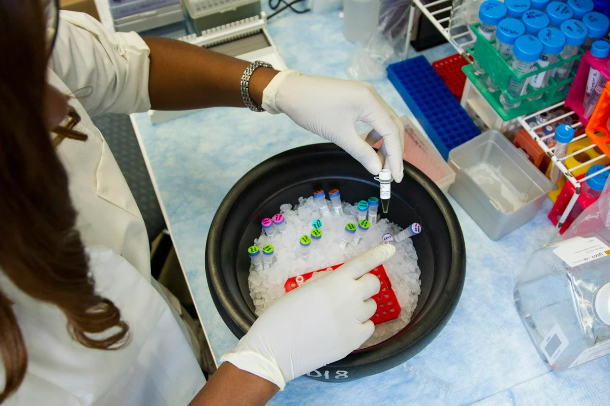
Illustrative photo · not a video frame
The Royal Society · 44 min 28 sec · Background explainer
Is this the AI future we want? | The Royal Society
YouTube loads only when you choose; playback starts when you press play.
A Royal Society discussion with Madhumita Murgia and biomedical engineer Alison Noble on AI’s potential in healthcare, science and culture, recorded at the 30 June 2026 event.
Evidence check: A discussion of opportunities and risks, including future possibilities. It is not evidence that every application described is available or clinically proven.
Published 2026-07-01. Source checked 14 October 2026. Check captions on the publisher’s video page.
Check the healthcare evidencePhoto: Daniel Sone / National Cancer Institute / Unsplash · Licence
Continue the story
Read next
Continue exploring machine learning: compare the finding and its evidence limits.
Can MRI machine learning detect focal cortical dysplasia?
A proof-of-concept study distinguished focal-cortical-dysplasia epilepsy from healthy controls with a highest internally cross-validated AUC of 0.791. There was no external clinical validation, and the model found only three of six MRI-negative cases.
Read the next reportExplore the full source and evidence record
What this means for people
- Children and adults could eventually receive an additional low-burden check from telemetry already collected during implant programming, but the method is not ready to replace imaging.
- Audiologists and clinical engineers would need a clear alert pathway that counts false alarms and keeps symptoms, hearing changes and device history visible.
- People with implants should not interpret a model estimate as a diagnosis or change follow-up based on this small retrospective study.
Global context
Cochlear-implant services differ in device mix, imaging access and follow-up capacity. A telemetry-based screen could be especially useful where repeated imaging is burdensome, but cross-manufacturer and cross-centre validation is essential before international adoption.
What the evidence does not yet show
- Only 32 evaluated implant cases: 16 confirmed migrations and 16 matched stable controls
- Case-control sampling with 50% migration prevalence cannot provide routine-clinic predictive values
- Retrospective and temporally asynchronous telemetry and imaging; no prospective alert timing
- MED-EL FLEX-series arrays only, with a training set dominated by one array type
- No hearing, quality-of-life, revision, radiation-reduction or cost outcome
- Manufacturer advisory roles and external grants disclosed by several authors
What to watch next
- Prospective screening in an unselected, low-prevalence clinic population
- External validation across manufacturers, array designs and telemetry systems
- Alert timing, false alarms per patient-year and missed migration consequences
- Hearing outcomes, imaging avoided, revision timing and patient experience
- Independent replication without device-company advisory or funding relationships
Living evidence record
Impact record IAI-0AW2KRA
Evidence stage
Studied
Confidence
Supported
Reporting basis
Source analysis
Independent or research support
Present
Record status
Monitoring
Last checked
4 November 2026
Source trail
1 direct source across 1 source type.
People impact
Documented in this record.
Uncertainty
Limits and next checks are explicit.
Stages describe the evidence available—not whether a technology is good or bad. See the public method.
Single-source reporting disclosure
This record analyses one direct source. It can establish what Journal of Medical Systems published or reported, but it is not independent corroboration of every performance claim or predicted outcome. The confidence label will change only when broader evidence is added.
Evidence trail
Sources used for this report
Links checked 4 November 2026
Research question, topics and regions
Whether routinely collected electrical impedance telemetry can identify postoperative cochlear-implant electrode-array migration without additional radiological imaging
Topics: Cochlear implants · Clinical monitoring · Medical devices · Machine learning · Paediatric care
Regions: Switzerland · Germany · Europe · Global
This report is labelled source analysis. We summarise and analyse source material in our own words; company statements remain attributed claims until independently supported. Translated summaries preserve the meaning of the original source and link back to it. Read our editorial standards.
Reader commentary
What does this mean where you are?
Tell us what you have seen at work, in your community or in everyday life. A question is welcome too. No account is needed; we review notes before publishing them.
Explore commentary across the portal →Published reader notes
0No published reader notes yet. You can start the evidence-led discussion above.
Prefer a private correction or response? Contact the newsroom.