Does an AI heart-MRI measurement predict survival better than clinicians?
In 27,382 UK and US patients, an automated ejection-fraction measurement was slightly more predictive of later mortality than the value recorded by clinicians. The study was retrospective and did not test treatment decisions, patient benefit or prospective deployment.
The short version
- What happened
- The retrospective study included 27,382 consecutive patients: 26,106 from a UK referral network and 1,276 from a US centre. During a median 5.6 years of follow-up, 3,458 patients died.
- Who it affects
- Patients could receive faster, more consistent first-pass measurements, but a result near a treatment threshold still needs clinical interpretation and visible quality control.
- What remains unproven
- The analysis was retrospective and cannot show that AI-guided measurement improves treatment, workload or survival.
Keep in mind
The analysis was retrospective and cannot show that AI-guided measurement improves treatment, workload or survival.
The cohort consisted of patients already referred for cardiac MRI, not a general or screening population.
Most participants came through one UK specialist-centre network; the US cohort was much smaller.
After age and sex adjustment, the discrimination advantage was statistically clear but only 0.004 in C-index terms.
About 1% of patients had a segmentation error, requiring a reliable review and escalation workflow.
Historical scans from 2010-2019 may not fully represent current scanners, protocols and treatment pathways.
The senior author is identified as the inventor of the AI tool; independent replication is needed.
Multi-source analysis · 2 linked sources. Primary analysed source date: 3 November 2026.
Keep following the evidence
Join The Impact Brief founding list for selected AI reports, their effect on people and the evidence limits. Read a sample first.
Read the full report · 9 min The evidence, methods, implications and limitations
The direct answer: yes, but the predictive gain became small after basic adjustment
The automated measurement was more strongly associated with later survival than the ejection-fraction value recorded by clinicians in this large retrospective dataset. In the unadjusted comparison, the AI measurement produced a C index of 0.669, compared with 0.637 for the clinician measurement. The difference was 0.033, with a 95% confidence interval from 0.026 to 0.041. A larger Akaike Information Criterion improvement also favoured the AI measurement. Those results mean the automated value ranked higher-risk patients somewhat better within the analysed cohort; they do not mean it predicted an individual's date or cause of death.
After the researchers added age and sex, both models discriminated more strongly and the gap narrowed sharply. The C index was 0.781 for the model using AI ejection fraction and 0.777 for the model using clinician ejection fraction, a difference of 0.004 with a 95% confidence interval from 0.002 to 0.006. That is statistically distinguishable in a dataset this large, but modest in practical terms. Whether it changes a treatment threshold, reduces missed disease or helps a patient cannot be inferred from a prognostic statistic alone.[1][2]
Who was studied and what outcome was counted
The investigators assembled 27,382 consecutive people who had cardiac MRI for known or suspected heart disease. The UK cohort contained 26,106 patients referred from nine hospitals to Barts Heart Centre between October 2014 and December 2019. A separate 1,276-patient cohort came from the University of Pittsburgh Medical Center between June 2010 and March 2016. These are clinical populations selected for advanced imaging, not a general-population screening sample.
The median follow-up was 5.6 years, with an interquartile range from 4.4 to 6.9 years. Mortality records showed 3,458 deaths, or 13% of the cohort. All-cause mortality is objective and important, but it is a broad endpoint: the study does not say whether deaths were cardiac, whether treatment after the scan differed, or whether an automated measurement would have changed care. It measures prognostic association from historical scans and records rather than the effect of deploying the tool.[1]
The distinction that matters
What the study compared—and what it did not test
The retrospective design compares measurements as predictors. It does not compare patient care with and without AI.
Cohort
27,382 cardiac-MRI patients in UK and US archives
3,458 deaths over a median 5.6 years
Comparison
AI-derived versus clinician-derived left-ventricular measurements
AI had slightly better mortality discrimination
Adjusted result
Age and sex added to the prognostic models
C index 0.781 versus 0.777
Not tested
No prospective AI-guided care pathway
Treatment, workload and patient benefit remain unproven
What the AI and clinician measurements were compared on
Ejection fraction describes the share of blood pumped from the left ventricle with each contraction. Clinicians use it when considering heart-failure medicines, implanted devices, procedures, surgical risk and monitoring during treatments such as chemotherapy. In cardiac MRI, the value depends on outlining the ventricle across images and calculating volumes. Small differences in those boundaries can alter the reported result, especially near a treatment threshold.
The researchers applied a deep-learning segmentation system to all scans and compared its ventricular volumes and ejection fraction with the measurements in the clinical archive. They fitted Cox proportional-hazards models and compared model fit, discrimination and the incremental information supplied when one measurement was added to the other. AI ejection fraction improved a model already containing the clinician value more than the clinician value improved a model already containing the AI result. That suggests the automated contours captured prognostic information more consistently, but it is not a randomized contest between AI-led and clinician-led care.[1]
Speed is the operational attraction—and segmentation errors define the safeguard
The university account says the system processed a scan in roughly two to three seconds. It contrasts that with previous work from the group estimating about 13 minutes for expert analysis. A consistent first-pass measurement could reduce repetitive contouring and make specialist time available for images that are difficult, unusual or close to a decision boundary. The present study did not directly measure staff time, waiting lists, costs or reporting turnaround after deployment, so those benefits remain plausible rather than demonstrated here.
Errors were uncommon but not absent. The institutional report puts the patient-level segmentation error rate at about 1%, affecting 0.08% of images. In a service processing thousands of scans, even a low percentage creates cases requiring recognition and correction. The paper's conclusion therefore pairs first-line automation with clinician oversight. A safe workflow needs a way to flag implausible contours, retain the source images, show what the model drew and route uncertain or threshold-adjacent cases for expert review rather than silently turning every output into a final result.[2]
What this could mean for patients and clinical teams
For patients, a faster and more reproducible ejection-fraction measurement could shorten the analytical part of a scan pathway and reduce variation between readers. The consequential question is whether that changes care appropriately. A value near a guideline threshold can affect medicine, device or follow-up decisions, so consistency may help. The same threshold sensitivity also raises risk: a confident but incorrect contour could move a person into or out of an intervention category unless the full clinical context is reviewed.
For imaging teams, the study supports evaluating an automated first pass rather than removing expertise. Services would need local validation across scanners, protocols, disease types and patient groups; a documented override route; and monitoring for drift or unusual failure modes. Clinicians should see the segmentation and relevant quality signals, not just a number. Patients should not be told that the system has proved it will extend survival: it predicted mortality slightly better in historical data, while the effect of using it in practice remains untested.[1][2]
Geography, funding and interests matter
The data came from UK referrals to one specialist centre and a much smaller US centre. That gives the analysis two health-system settings, but the sample is heavily weighted toward one UK network and historical imaging from 2010 to 2019. Scanner hardware, reporting practice and treatment pathways have changed. Performance should not be assumed in lower-resource services, community imaging, paediatric care or populations not well represented among patients sent for cardiac MRI.
The work was funded through National Institute for Health and Care Research support, the NIHR UCLH Biomedical Research Centre and a British Heart Foundation Accelerator Award. UCL identifies the senior author as the inventor of the AI tool. That interest does not invalidate the results, but it strengthens the case for independent replication, transparent software versioning and prospective evaluation by services that did not create the system.[1][2]
Evidence check and what would change the assessment
This is peer-reviewed, multicentre evidence with a large consecutive cohort, long follow-up and 3,458 outcome events. The comparison uses established survival-analysis measures and reports uncertainty for the discrimination difference. Those are substantial strengths. Its central limitation is design: retrospective prediction cannot establish that introducing the tool improves decisions, reduces workload safely or changes survival. The model was assessed on patients who already reached cardiac MRI, so it also says nothing about access to scanning or earlier diagnostic bottlenecks.
Confidence in routine first-line use would rise with a prospective, preferably independently led study that prespecifies failure rules and tests a real workflow. It should compare reporting time, corrections, threshold reclassification, downstream testing, treatment decisions, clinician workload and patient outcomes—not only another C index. Results should be stratified by centre, scanner, sex, age, ethnicity and important cardiac conditions. If automated measurement remains accurate under those conditions, catches its own failures and improves care without widening access gaps, the practical case would be much stronger. If its small adjusted prognostic advantage disappears or creates harmful reclassification, the appropriate role may remain decision support for selected scans.[1][2]
Watch the background, then check the evidence
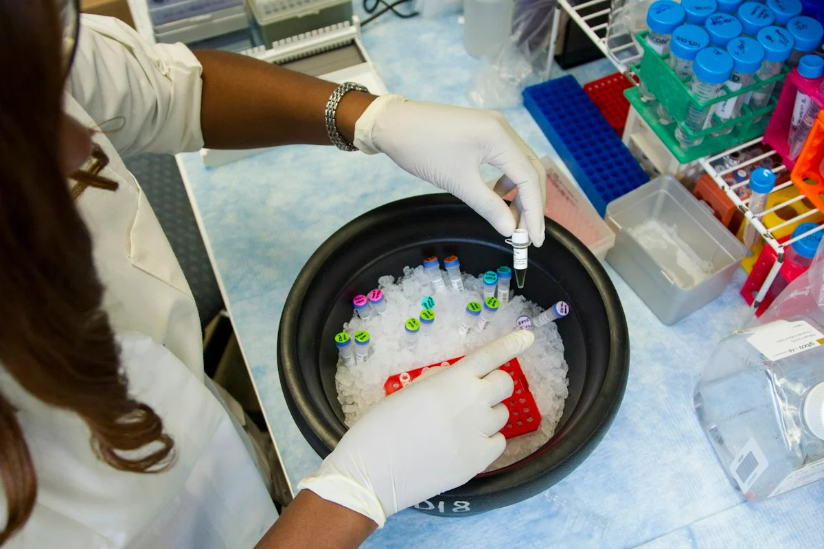
Illustrative photo · not a video frame
The Royal Society · 44 min 28 sec · Background explainer
Is this the AI future we want? | The Royal Society
YouTube loads only when you choose; playback starts when you press play.
A Royal Society discussion with Madhumita Murgia and biomedical engineer Alison Noble on AI’s potential in healthcare, science and culture, recorded at the 30 June 2026 event.
Evidence check: A discussion of opportunities and risks, including future possibilities. It is not evidence that every application described is available or clinically proven.
Published 2026-07-01. Source checked 14 October 2026. Check captions on the publisher’s video page.
Check the healthcare evidencePhoto: Daniel Sone / National Cancer Institute / Unsplash · Licence
Continue the story
Read next
Continue exploring medical imaging: compare the finding and its evidence limits.
Can MRI machine learning detect focal cortical dysplasia?
A proof-of-concept study distinguished focal-cortical-dysplasia epilepsy from healthy controls with a highest internally cross-validated AUC of 0.791. There was no external clinical validation, and the model found only three of six MRI-negative cases.
Read the next reportExplore the full source and evidence record
What this means for people
- Patients could receive faster, more consistent first-pass measurements, but a result near a treatment threshold still needs clinical interpretation and visible quality control.
- Imaging teams may save contouring time if the system performs locally; the study did not measure real service workload or turnaround after deployment.
- Health systems need independent validation because most patients came through one UK specialist network and the tool's inventor was part of the research team.
Global context
The study spans UK and US specialist cardiac-MRI services but is dominated by one UK referral network. Cardiac imaging access, scanner protocols, staffing and treatment thresholds differ internationally. A fast automated measurement may be especially attractive where expert time is scarce, yet importing a model without local calibration and an override pathway could transfer errors into settings least able to absorb them.
What the evidence does not yet show
- The analysis was retrospective and cannot show that AI-guided measurement improves treatment, workload or survival.
- The cohort consisted of patients already referred for cardiac MRI, not a general or screening population.
- Most participants came through one UK specialist-centre network; the US cohort was much smaller.
- After age and sex adjustment, the discrimination advantage was statistically clear but only 0.004 in C-index terms.
- About 1% of patients had a segmentation error, requiring a reliable review and escalation workflow.
- Historical scans from 2010-2019 may not fully represent current scanners, protocols and treatment pathways.
- The senior author is identified as the inventor of the AI tool; independent replication is needed.
What to watch next
- Prospective multicentre deployment with prespecified failure and override rules
- Effects on reporting time, workload, treatment decisions and patient outcomes
- Performance across scanners, centres, demographic groups and cardiac conditions
- Independent replication and transparent handling of inventor or intellectual-property interests
- Monitoring of threshold reclassification and the roughly 1% patient-level error rate
Living evidence record
Impact record IAI-0DT1IK9
Evidence stage
Studied
Confidence
Supported
Reporting basis
Multi-source analysis
Independent or research support
Present
Record status
Monitoring
Last checked
4 November 2026
Source trail
2 direct sources across 2 source types.
People impact
Documented in this record.
Uncertainty
Limits and next checks are explicit.
Stages describe the evidence available—not whether a technology is good or bad. See the public method.
Evidence trail
Sources used for this report
Links checked 4 November 2026
Research question, topics and regions
Whether AI-derived left-ventricular measurements from cardiac MRI predict all-cause mortality more accurately than measurements recorded by clinicians
Topics: Cardiac MRI · Ejection fraction · Prognostic modelling · Clinical oversight · Medical imaging
Regions: United Kingdom · United States · Europe · North America
This report is labelled multi-source analysis. We summarise and analyse source material in our own words; company statements remain attributed claims until independently supported. Translated summaries preserve the meaning of the original source and link back to it. Read our editorial standards.
Reader commentary
What does this mean where you are?
Tell us what you have seen at work, in your community or in everyday life. A question is welcome too. No account is needed; we review notes before publishing them.
Explore commentary across the portal →Published reader notes
0No published reader notes yet. You can start the evidence-led discussion above.
Prefer a private correction or response? Contact the newsroom.