Can an MRI model predict cancer progression? A 33-patient holdout is promising, not treatment guidance
A Chinese single-centre model separated progression risk after chemoradiotherapy in 113 patients, but the holdout contained only 33 people and no external hospital. The paper's feature count is also internally inconsistent.
The short version
- What happened
- Researchers retrospectively analysed 113 patients from one Shandong hospital: 80 for model development and 33 for a held-out internal test.
- Who it affects
- People facing a rare head-and-neck cancer need better estimates of progression risk, but a model should not determine treatment intensity or follow-up until another hospital can reproduce the predictions and show that using them improves decisions without avoidable harm.
- What remains unproven
- Retrospective single-centre cohort
Keep in mind
Retrospective single-centre cohort
113 patients total
33-patient internal holdout
No external validation
No prospective decision test
One MRI sequence and scanner context
Feature-count inconsistency in the paper
No demonstrated patient benefit
Source analysis · 1 linked source. Primary analysed source date: 28 October 2026.
Keep following the evidence
Join The Impact Brief founding list for selected AI reports, their effect on people and the evidence limits. Read a sample first.
Read the full report · 3 min The evidence, methods, implications and limitations
The model found a signal in pretreatment MRI
Researchers used records from 113 people with hypopharyngeal squamous-cell cancer treated with concurrent chemoradiotherapy at one Shandong hospital between 2016 and 2019. They developed the model in 80 patients and reserved 33 for a final internal test. Two experienced radiologists outlined tumours on diffusion-weighted MRI, from which the team extracted 1,288 image features.
A radiomics score alone produced validation AUCs of 0.737 for one-year and 0.715 for two-year progression-free survival. Combining the score with age and tumour T stage yielded a reported validation C-index of 0.858, but the confidence interval was 0.690 to 1.000. High- and low-risk groups also differed in the holdout set at p=0.023.[1]
The study cannot support changing treatment or follow-up
The holdout came from the same hospital, scanner family and historical care pathway as the training data. Thirty-three patients are too few to establish calibration, subgroup performance or transport to another clinic. The study did not compare model-guided care with usual practice and measured no benefit from adding immunotherapy, intensifying chemotherapy or changing scan intervals.
Reproducibility also needs clarification: the abstract and results say six radiomic features were selected, while the published coefficient table lists seven. The authors acknowledge the single-centre, retrospective design, small sample and use of one MRI sequence. A preregistered external validation with locked code and treatment-independent evaluation would change the assessment.[1]
The distinction that matters
What was tested—and what was not
The paper tests prediction inside one historical dataset, not a clinical decision pathway.
Develop
Select features in 80 patients
Training-only preprocessing and cutoff
Hold out
Test in 33 patients from the same hospital
C-index 0.858; wide uncertainty
Missing
Use the score to change care prospectively
No treatment or patient-outcome evidence
Watch the background, then check the evidence
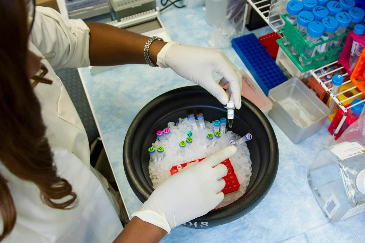
Illustrative photo · not a video frame
The Royal Society · 44 min 28 sec · Background explainer
Is this the AI future we want? | The Royal Society
YouTube loads only when you choose; playback starts when you press play.
A Royal Society discussion with Madhumita Murgia and biomedical engineer Alison Noble on AI’s potential in healthcare, science and culture, recorded at the 30 June 2026 event.
Evidence check: A discussion of opportunities and risks, including future possibilities. It is not evidence that every application described is available or clinically proven.
Published 2026-07-01. Source checked 14 October 2026. Check captions on the publisher’s video page.
Check the healthcare evidencePhoto: Daniel Sone / National Cancer Institute / Unsplash · Licence
Continue the story
Read next
Continue exploring clinical ai: compare the finding and its evidence limits.
Cancer-fever model travelled to a second hospital—but still needs a treatment trial
A model developed on 987 German hospital stays and tested on 7,409 US stays predicted whether fever would persist after antibiotics, with external AUROC 0.78. Retrospective opportunity counts do not show that withholding escalation is safe.
Read the next reportExplore the full source and evidence record
What this means for people
- People facing a rare head-and-neck cancer need better estimates of progression risk, but a model should not determine treatment intensity or follow-up until another hospital can reproduce the predictions and show that using them improves decisions without avoidable harm.
Global context
The cohort came from one Chinese hospital and one treatment period. Scanner settings, tumour mix, treatment practice and follow-up differ across health systems, so the reported score cannot be assumed to travel.
What the evidence does not yet show
- Retrospective single-centre cohort
- 113 patients total
- 33-patient internal holdout
- No external validation
- No prospective decision test
- One MRI sequence and scanner context
- Feature-count inconsistency in the paper
- No demonstrated patient benefit
What to watch next
- Locked external validation
- Prospective multi-centre evaluation
- Clarification of the six-versus-seven feature count
- Calibration across scanners and hospitals
- Decision-impact and safety outcomes
- Open model code and reproducible preprocessing
Living evidence record
Impact record IAI-1LCYQP3
Evidence stage
Studied
Confidence
Supported
Reporting basis
Source analysis
Independent or research support
Present
Record status
Monitoring
Last checked
28 October 2026
Source trail
1 direct source across 1 source type.
People impact
Documented in this record.
Uncertainty
Limits and next checks are explicit.
Stages describe the evidence available—not whether a technology is good or bad. See the public method.
Single-source reporting disclosure
This record analyses one direct source. It can establish what Frontiers in Oncology published or reported, but it is not independent corroboration of every performance claim or predicted outcome. The confidence label will change only when broader evidence is added.
Evidence trail
Sources used for this report
Links checked 28 October 2026
Research question, topics and regions
Whether texture features from pretreatment diffusion-weighted MRI can add progression-risk information for people with hypopharyngeal cancer receiving concurrent chemoradiotherapy
Topics: Clinical AI · Cancer · MRI · Radiomics · Prognosis · External validation
Regions: China · Asia
This report is labelled source analysis. We summarise and analyse source material in our own words; company statements remain attributed claims until independently supported. Translated summaries preserve the meaning of the original source and link back to it. Read our editorial standards.
Reader commentary
What does this mean where you are?
Tell us what you have seen at work, in your community or in everyday life. A question is welcome too. No account is needed; we review notes before publishing them.
Explore commentary across the portal →Published reader notes
0No published reader notes yet. You can start the evidence-led discussion above.
Prefer a private correction or response? Contact the newsroom.