Ear-disease AI narrowed the trainee–expert gap—but also changed 46 correct answers to wrong ones
Thirteen clinicians re-read 100 disease-enriched ear images with AI advice. Accuracy rose most for junior residents, but the single-centre retrospective study cannot show how the tool performs in routine care.
The short version
- What happened
- A South Korean model trained on 7,335 otoendoscopy images was tested with three professors, five senior residents and five junior residents on an independent 100-image set.
- Who it affects
- A less experienced clinician may gain a useful second opinion from image support, but a confident model answer can also pull a correct judgement off course. Patients need prospective evidence that the combined clinician–AI workflow improves real diagnoses and care.
- What remains unproven
- Single-centre retrospective study using one South Korean image database and a 100-image assessment set.
Keep in mind
Single-centre retrospective study using one South Korean image database and a 100-image assessment set.
Disease-enriched set excluded fully normal images and cases with discordant annotations, limiting routine-care generalisability.
The same clinicians read the images twice, three months apart; intrareader variability may explain some changes.
No prospective workflow, referral, treatment or patient outcome was measured.
Funded by the Korean Ministry of Food and Drug Safety; the authors did not disclose competing interests in the publisher record.
Source analysis · 1 linked source. Primary analysed source date: 30 October 2026.
Keep following the evidence
Join The Impact Brief founding list for selected AI reports, their effect on people and the evidence limits. Read a sample first.
Read the full report · 3 min The evidence, methods, implications and limitations
Trainees improved most on the same 100 images
AI advice substantially raised image-classification accuracy in a peer-reviewed South Korean reader study. Three otology professors, five senior residents and five junior residents first classified 100 otoendoscopy images without assistance, then repeated the task with model predictions three months later. For the main categories—chronic otitis media, otitis media with effusion or neither—accuracy rose by 11.0 percentage points for professors, 28.8 points for senior residents and 32.2 points for junior residents.
The model had been trained on 7,335 images from Asan Medical Center. On the 100-image assessment set, its stand-alone primary-class accuracy was 95.0%. The gap between professors and junior residents narrowed from 24.3 to 3.1 percentage points. That is useful evidence about decision support under a one-minute-per-image constraint, not proof of faster or safer patient care.[1]
AI corrected many errors and introduced some
The paired decisions show both sides of assistance. Across 1,300 primary-class assessments, clinicians' first answer differed from the AI recommendation 499 times. They accepted the recommendation in 395 of those cases. Overall, support corrected 384 initial errors, while 46 initially correct decisions became incorrect. Junior residents were especially likely to accept AI advice and were least often right when they rejected it.
The assessment deliberately excluded fully normal images and therefore did not reflect the prevalence seen in ordinary clinics. Difficult cases with discordant annotations were also removed, the readers saw the same images twice, and all training data came from one centre and endoscope setting. Multicentre prospective studies should measure diagnostic errors, referrals, treatment and patient outcomes before broad deployment.[1]
The distinction that matters
What changed when clinicians saw the model's answer
Each of 13 clinicians classified the same 100 images before and after AI advice.
Junior residents
54.4% to 86.6%
Largest primary-class accuracy gain
Senior residents
65.0% to 93.8%
Highest assisted group accuracy
All readers
384 errors fixed; 46 errors introduced
Help and automation risk appeared together
Watch the background, then check the evidence
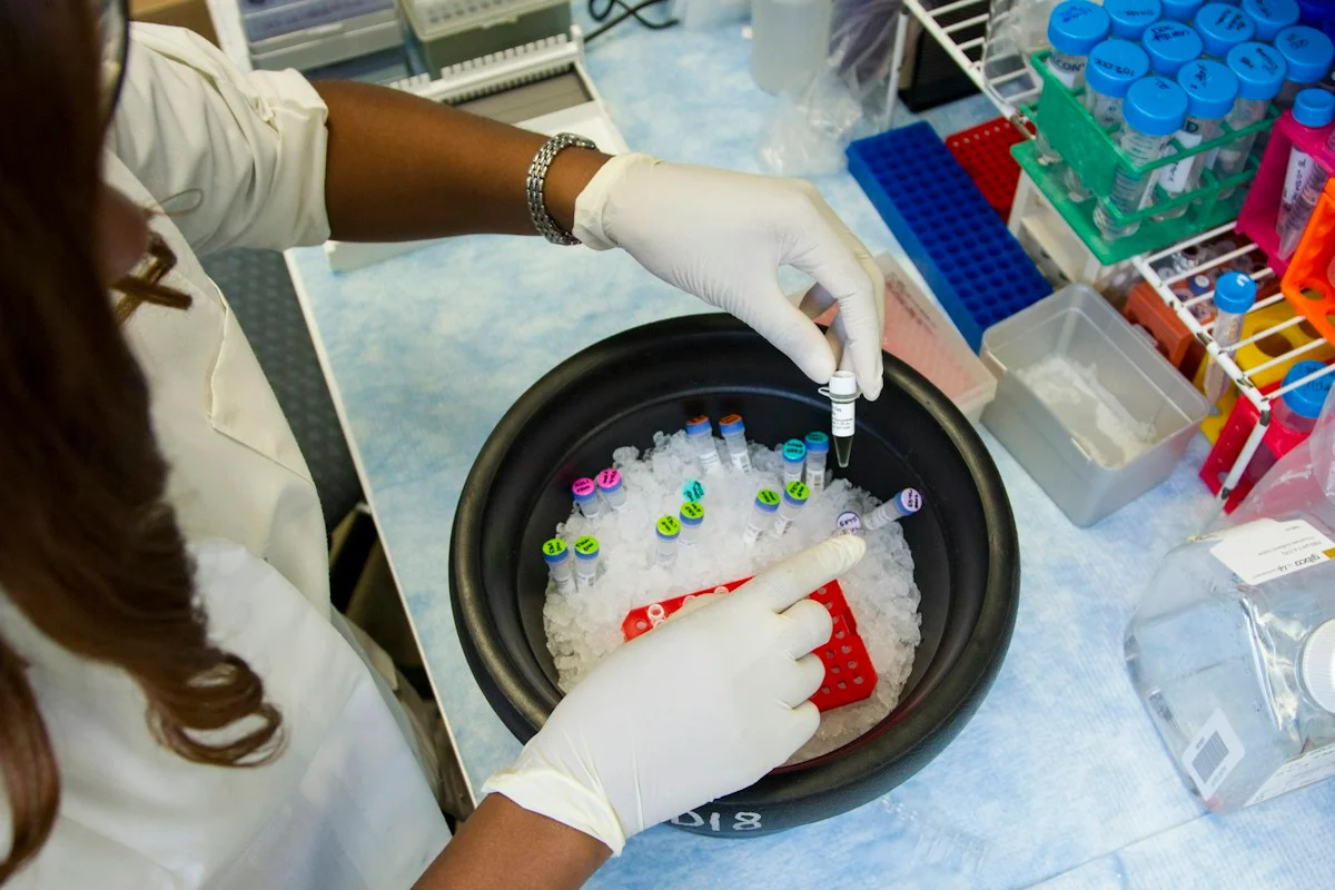
Illustrative photo · not a video frame
The Royal Society · 44 min 28 sec · Background explainer
Is this the AI future we want? | The Royal Society
YouTube loads only when you choose; playback starts when you press play.
A Royal Society discussion with Madhumita Murgia and biomedical engineer Alison Noble on AI’s potential in healthcare, science and culture, recorded at the 30 June 2026 event.
Evidence check: A discussion of opportunities and risks, including future possibilities. It is not evidence that every application described is available or clinically proven.
Published 2026-07-01. Source checked 14 October 2026. Check captions on the publisher’s video page.
Check the healthcare evidencePhoto: Daniel Sone / National Cancer Institute / Unsplash · Licence
Continue the story
Read next
Continue exploring automation bias: compare the finding and its evidence limits.
Can paediatricians resist wrong AI advice?
A Greek study of 98 paediatricians found that doctors usually rejected deliberately incorrect advice labelled as AI-generated, but residents and private-practice clinicians were more likely than hospital attendings to change towards a wrong answer. The experiment used fabricated advice and fixed-order vignettes, not a live clinical system.
Read the next reportExplore the full source and evidence record
What this means for people
- A less experienced clinician may gain a useful second opinion from image support, but a confident model answer can also pull a correct judgement off course. Patients need prospective evidence that the combined clinician–AI workflow improves real diagnoses and care.
Global context
Specialist shortages make ear-image decision support attractive beyond South Korea, but differences in patient mix, image devices, disease prevalence and referral pathways mean the reported accuracy cannot be transferred unchanged.
What the evidence does not yet show
- Single-centre retrospective study using one South Korean image database and a 100-image assessment set.
- Disease-enriched set excluded fully normal images and cases with discordant annotations, limiting routine-care generalisability.
- The same clinicians read the images twice, three months apart; intrareader variability may explain some changes.
- No prospective workflow, referral, treatment or patient outcome was measured.
- Funded by the Korean Ministry of Food and Drug Safety; the authors did not disclose competing interests in the publisher record.
What to watch next
- Multicentre prospective validation
- Performance with normal and difficult images
- Calibration and visible uncertainty
- Automation-bias training for clinicians
- Patient-level diagnostic and treatment outcomes
- External validation across devices and regions
Living evidence record
Impact record IAI-0JR20DY
Evidence stage
Studied
Confidence
Supported
Reporting basis
Source analysis
Independent or research support
Present
Record status
Monitoring
Last checked
31 October 2026
Source trail
1 direct source across 1 source type.
People impact
Documented in this record.
Uncertainty
Limits and next checks are explicit.
Stages describe the evidence available—not whether a technology is good or bad. See the public method.
Single-source reporting disclosure
This record analyses one direct source. It can establish what JMIR Medical Informatics published or reported, but it is not independent corroboration of every performance claim or predicted outcome. The confidence label will change only when broader evidence is added.
Evidence trail
Sources used for this report
Links checked 31 October 2026
Research question, topics and regions
Whether AI advice improves clinicians' classification of middle-ear disease across experience levels
Topics: Clinical AI · Ear disease · Medical imaging · Automation bias · Medical education
Regions: Asia · South Korea · Global
This report is labelled source analysis. We summarise and analyse source material in our own words; company statements remain attributed claims until independently supported. Translated summaries preserve the meaning of the original source and link back to it. Read our editorial standards.
Reader commentary
What does this mean where you are?
Tell us what you have seen at work, in your community or in everyday life. A question is welcome too. No account is needed; we review notes before publishing them.
Explore commentary across the portal →Published reader notes
0No published reader notes yet. You can start the evidence-led discussion above.
Prefer a private correction or response? Contact the newsroom.