Synthetic radiomic features improved throat-lesion classification—but only 30 patients tested the claim
A generative model helped a classifier distinguish benign from malignant laryngeal lesions in one retrospective dataset. The held-out test was small and no external hospital or clinical workflow was evaluated.
The short version
- What happened
- The retrospective dataset contained 11,144 narrow-band images from 210 adults; 180 patients were used for development and 30 for an internal held-out test.
- Who it affects
- Earlier, more consistent recognition of suspicious throat lesions could shorten diagnosis, but the published experiment cannot show that this model changes biopsy decisions or outcomes.
- What remains unproven
- Only 30 patients in the held-out test
Keep in mind
Only 30 patients in the held-out test
Single retrospective data source
No external-centre validation
No clinician, workflow or patient-outcome comparison
Source analysis · 1 linked source. Primary analysed source date: 24 October 2026.
Keep following the evidence
Join The Impact Brief founding list for selected AI reports, their effect on people and the evidence limits. Read a sample first.
Read the full report · 2 min The evidence, methods, implications and limitations
The experiment augmented measurements rather than inventing clinical pictures
The researchers analysed 11,144 narrow-band endoscopy images from 210 adults with benign or malignant laryngeal lesions. They extracted numerical radiomic measurements, then trained generative adversarial networks to create extra feature vectors for under-represented classes. The generated material was therefore tabular data derived from images, not synthetic endoscopy photographs.
Development used 180 patients and an internal held-out test used 30. Across repeated experiments, the best generative pipeline recorded a median F1 score of 0.851, versus 0.806 for the conventional SMOTE oversampling baseline. F1 combines precision and recall; it does not by itself show how often a clinician would miss a cancer or recommend an unnecessary biopsy in practice.[1]
Thirty held-out patients cannot establish clinical readiness
The patient-level split is important because images from the same person should not leak across training and testing. Even so, a 30-patient test from the same retrospective source is too small to establish transportability. The paper does not report a prospective clinician comparison, workflow effects or patient outcomes.
A stronger assessment would freeze the full pipeline and test consecutive patients at independent centres, reporting sensitivity and specificity with confidence intervals by lesion type and image quality. It should also compare specialists with and without support and track biopsies, delays and missed disease. The authors reported no dedicated external funding and no competing interests.[1]
The distinction that matters
What was generated and what was tested
The study augmented extracted radiomic measurements before classifying lesions.
Source data
11,144 images from 210 adults
Extract radiomic features
Generative step
Create numerical feature vectors
Balance development data
Held-out test
30 patients
Median F1 0.851
Watch the background, then check the evidence
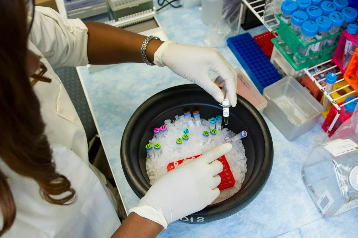
Illustrative photo · not a video frame
The Royal Society · 44 min 28 sec · Background explainer
Is this the AI future we want? | The Royal Society
YouTube loads only when you choose; playback starts when you press play.
A Royal Society discussion with Madhumita Murgia and biomedical engineer Alison Noble on AI’s potential in healthcare, science and culture, recorded at the 30 June 2026 event.
Evidence check: A discussion of opportunities and risks, including future possibilities. It is not evidence that every application described is available or clinically proven.
Published 2026-07-01. Source checked 14 October 2026. Check captions on the publisher’s video page.
Check the healthcare evidencePhoto: Daniel Sone / National Cancer Institute / Unsplash · Licence
Continue the story
Read next
Continue exploring medical imaging: compare the finding and its evidence limits.
Can generative imaging rescue a poorly timed stroke scan?
Adding generated vessel images improved four readers’ large-vessel-occlusion detection in a 194-case simulated test. The real failed-scan cohort had only 41 patients and two target occlusions, and the model sometimes omitted vessels without an obvious artefact.
Read the next reportExplore the full source and evidence record
What this means for people
- Earlier, more consistent recognition of suspicious throat lesions could shorten diagnosis, but the published experiment cannot show that this model changes biopsy decisions or outcomes.
Global context
Endoscopy equipment, disease prevalence and referral practice vary across health systems, making independent multi-centre validation essential.
What the evidence does not yet show
- Only 30 patients in the held-out test
- Single retrospective data source
- No external-centre validation
- No clinician, workflow or patient-outcome comparison
What to watch next
- Locked external validation
- Per-lesion sensitivity and false-positive rates
- Specialist reader study
- Prospective biopsy and diagnosis outcomes
Living evidence record
Impact record IAI-0TZAEFK
Evidence stage
Studied
Confidence
Supported
Reporting basis
Source analysis
Independent or research support
Present
Record status
Monitoring
Last checked
25 October 2026
Source trail
1 direct source across 1 source type.
People impact
Documented in this record.
Uncertainty
Limits and next checks are explicit.
Stages describe the evidence available—not whether a technology is good or bad. See the public method.
Single-source reporting disclosure
This record analyses one direct source. It can establish what BMC Medical Imaging published or reported, but it is not independent corroboration of every performance claim or predicted outcome. The confidence label will change only when broader evidence is added.
Evidence trail
Sources used for this report
Links checked 25 October 2026
Research question, topics and regions
Whether synthetic radiomic feature vectors improve laryngeal-lesion classification from narrow-band imaging
Topics: Laryngeal cancer · Radiomics · Medical imaging · Generative AI
Regions: Italy · Europe · Global
This report is labelled source analysis. We summarise and analyse source material in our own words; company statements remain attributed claims until independently supported. Translated summaries preserve the meaning of the original source and link back to it. Read our editorial standards.
Reader commentary
What does this mean where you are?
Tell us what you have seen at work, in your community or in everyday life. A question is welcome too. No account is needed; we review notes before publishing them.
Explore commentary across the portal →Published reader notes
0No published reader notes yet. You can start the evidence-led discussion above.
Prefer a private correction or response? Contact the newsroom.