Synthetic rare-lesion images helped five novice endoscopists—what remains unproven
A peer-reviewed study found better image-level recognition after novices trained on generated examples, but the same fixed test was used before and after and no patient outcomes were measured.
The short version
- What happened
- Five novice endoscopists improved on a fixed image-recognition test after seeing synthetic examples of four rare gastrointestinal lesions; the study did not test real procedures or patient outcomes.
- Who it affects
- Novice clinicians could gain more practice with conditions they rarely encounter, but realistic synthetic images can also create false confidence if training performance is mistaken for readiness to diagnose patients.
- What remains unproven
- The reader study included five novices, four conditions and the same fixed mixed image set before and after synthetic exposure, with no separate control group.
Keep in mind
The reader study included five novices, four conditions and the same fixed mixed image set before and after synthetic exposure, with no separate control group.
The classifier results came from curated image datasets and do not establish performance across hospitals, devices, prevalence levels or live procedures.
Two conditions showed ceiling effects, limiting the ability to detect improvement.
The approach reduces fine-tuning on patient pixels but has not undergone the formal privacy audits that the authors recommend.
Source analysis · 2 linked sources. Primary analysed source date: 25 October 2026. Earliest linked supporting source: 30 December 2025.
Keep following the evidence
Join The Impact Brief founding list for selected AI reports, their effect on people and the evidence limits. Read a sample first.
Read the full report · 4 min The evidence, methods, implications and limitations

Generated cases improved a narrow training exercise
Researchers led from Fudan University built EndoRare to generate varied endoscopy images from one real reference image for each of four uncommon conditions: calcifying fibrous tumour, juvenile polyposis syndrome, familial adenomatous polyposis and Peutz–Jeghers syndrome. Five novice endoscopists first studied one real anchor image per condition, took a fixed mixed-image test, then saw generated examples and repeated that same test. Overall recall rose by 0.400 and precision by 0.267. Gains were largest for the two less distinctive-looking conditions; performance on the other two was already near the ceiling.
That is useful evidence for an educational aid, not proof of safer diagnosis. A five-person before-and-after exercise can reflect familiarity with the repeated test, and image recognition is only one part of endoscopy. The study did not compare synthetic training with a separate control curriculum, follow retention over time or measure decisions during live procedures. Clinicians and training programmes should therefore treat the images as supplementary practice material, not substitute patients or validated competency assessment.[1][2]
The model results are promising, with important limits
In a blinded assessment of 700 randomly sampled images on a three-point scale, EndoRare received the highest overall rating, 2.32, compared with 2.09 for fine-tuned Stable Diffusion and 2.01 for DreamBooth; inter-rater agreement was moderate-to-substantial at Fleiss' kappa 0.59. A rare-lesion classifier trained with EndoRare augmentation reached macro precision–recall AUC 0.708, against 0.612 for Textual Inversion, and macro ROC-AUC 0.978. These are image-test metrics, not diagnostic accuracy in a hospital population.
The authors froze the diffusion backbone instead of fine-tuning it on the rare reference image, which may reduce direct copying. They explicitly say that this does not eliminate privacy risk and call for nearest-neighbour, membership-inference and attribute-leakage tests. Confidence would rise with a larger, multi-centre randomised reader study using unseen test cases, multiple devices and experience levels, retention follow-up, independent privacy testing and prospective evaluation during real clinical work.[1][2]
The distinction that matters
What the evidence actually follows
The paper tests three different steps; only the first two are model or image outcomes.
Generate
Create varied examples from one rare-lesion reference
Images rated for realism and class faithfulness
Train
Augment a classifier and expose five novices to synthetic cases
Higher image-test metrics in the reported experiments
Validate
Run controlled, prospective multi-centre studies
Still needed before claiming clinical benefit
Watch the background, then check the evidence
Illustrative photo · not a video frame
The Royal Society · 44 min 28 sec · Background explainer
Is this the AI future we want? | The Royal Society
YouTube loads only when you choose; playback starts when you press play.
A Royal Society discussion with Madhumita Murgia and biomedical engineer Alison Noble on AI’s potential in healthcare, science and culture, recorded at the 30 June 2026 event.
Evidence check: A discussion of opportunities and risks, including future possibilities. It is not evidence that every application described is available or clinically proven.
Published 2026-07-01. Source checked 14 October 2026. Check captions on the publisher’s video page.
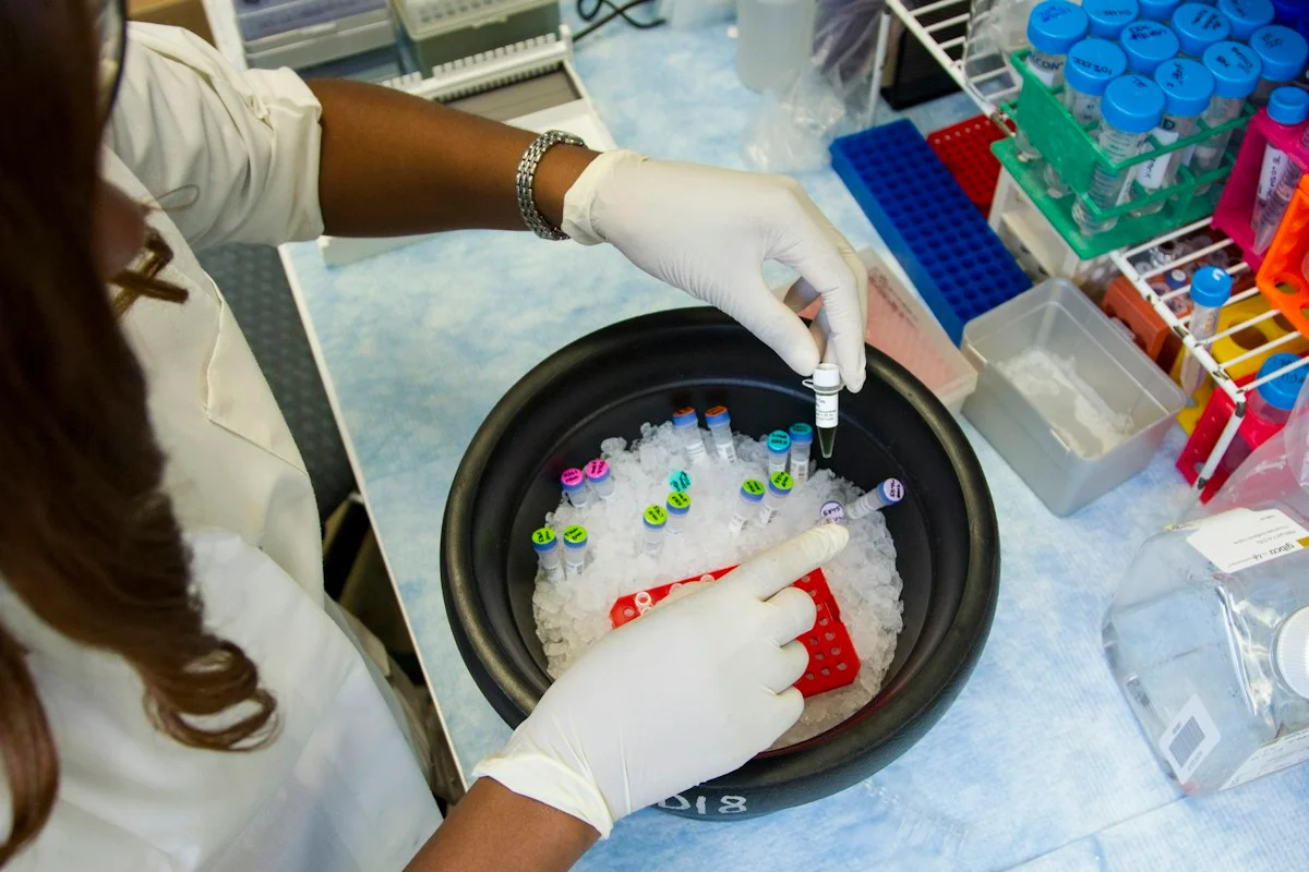
Check the healthcare evidencePhoto: Daniel Sone / National Cancer Institute / Unsplash · Licence
Continue the story
Read next
Continue exploring medical imaging: compare the finding and its evidence limits.
Can an adult X-ray AI learn neonatal care without moving patient data?
Across 30 neonatal intensive-care units, sequential local retraining cut average anatomical landmark error to 0.67 cm, compared with 1.29 cm for one-hospital fine-tuning and 4.30 cm for the unadapted adult model. The retrospective study did not deploy the system or measure harm, treatment changes or infant outcomes.
Read the next reportExplore the full source and evidence record
What this means for people
- Novice clinicians could gain more practice with conditions they rarely encounter, but realistic synthetic images can also create false confidence if training performance is mistaken for readiness to diagnose patients.
Global context
Rare-disease training data are scarce in every health system, but this evidence comes mainly from a Shanghai-centred research collaboration. Visual protocols, devices, populations and governance differ across hospitals, so local validation is essential.
What the evidence does not yet show
- The reader study included five novices, four conditions and the same fixed mixed image set before and after synthetic exposure, with no separate control group.
- The classifier results came from curated image datasets and do not establish performance across hospitals, devices, prevalence levels or live procedures.
- Two conditions showed ceiling effects, limiting the ability to detect improvement.
- The approach reduces fine-tuning on patient pixels but has not undergone the formal privacy audits that the authors recommend.
What to watch next
- Randomised multi-centre reader studies with unseen cases and retention follow-up
- Prospective evaluation during clinical workflows rather than image-only tests
- Independent privacy, memorisation and attribute-leakage audits
- Performance across devices, hospitals, experience levels and realistic prevalence
Living evidence record
Impact record IAI-0L1VIQM
Evidence stage
Studied
Confidence
Supported
Reporting basis
Source analysis
Independent or research support
Present
Record status
Monitoring
Last checked
25 October 2026
Source trail
2 direct sources across 1 source type.
People impact
Documented in this record.
Uncertainty
Limits and next checks are explicit.
Stages describe the evidence available—not whether a technology is good or bad. See the public method.
Related-source reporting disclosure
This record analyses 2 linked source records around the same underlying development. The extra records add method, date or context, but they do not by themselves constitute independent replication of every performance claim or predicted outcome.
Evidence trail
Sources used for this report
Links checked 25 October 2026
Research question, topics and regions
Whether one-shot synthetic endoscopy images can improve rare-lesion classifiers and novice image recognition without implying proven clinical benefit
Topics: Medical imaging · Clinical education · Synthetic data · Rare disease · Privacy
Regions: China · United Kingdom
This report is labelled source analysis. We summarise and analyse source material in our own words; company statements remain attributed claims until independently supported. Translated summaries preserve the meaning of the original source and link back to it. Read our editorial standards.
Reader commentary
What does this mean where you are?
Tell us what you have seen at work, in your community or in everyday life. A question is welcome too. No account is needed; we review notes before publishing them.
Explore commentary across the portal →Published reader notes
0No published reader notes yet. You can start the evidence-led discussion above.
Prefer a private correction or response? Contact the newsroom.