Routine scans revealed liver-cancer survival signals—but not a treatment rule
AI measured nine abdominal organs in 1,512 people receiving immunotherapy for liver cancer. Three continuous measurements tracked overall survival after correction, but the retrospective associations do not show that organ size changes treatment benefit.
The short version
3 key points
- What changed
- The retrospective study analysed 1,512 patients with hepatocellular carcinoma across 10 cohorts at six Chinese centres; 986 died and 1,298 progressed or died during follow-up.
- Why it matters
- Patients could eventually gain more prognostic information from scans they already undergo, but premature use could wrongly change a cancer treatment. The study measured associations, not better treatment choices or quality of life.
- What to watch
- Prospective multinational validation, paired scan-method comparisons and decision-impact studies showing whether organ features add useful information to established liver-cancer prognostic tools.
AI extracted more information from scans already taken
Researchers used a pretrained deep-learning system to segment nine abdominal organs or structures on pretreatment CT or MRI scans. The 1,512 adults came from 10 cohorts at six Chinese centres; 84.1% were men, 80.8% had advanced BCLC stage C disease and 82.5% had hepatitis B. Median follow-up was 32.03 months.[1]
Why the result is not a clinical decision rule
Three continuous measures remained associated with overall survival: larger liver volume and left-adrenal volume tracked lower mortality, while a larger height-normalised right kidney tracked higher mortality. No continuous measure survived correction for progression-free survival. In a separate threshold analysis, no organ measure retained a corrected association with overall survival.[1]
Who it affects
Patients could eventually gain more prognostic information from scans they already undergo, but premature use could wrongly change a cancer treatment. The study measured associations, not better treatment choices or quality of life.[1]
Keep in mind
Retrospective Chinese cohorts; 84.1% male and 82.5% hepatitis-B positive; residual confounding; inconsistent treatment-line data; locally assessed progression; unpaired CT and MRI; no prospective test of added clinical value.
Source analysis · 1 linked source. Original source date: 17 October 2026.
Continue the story
Read next
Health & Life Sciences
A lung-nodule model travelled better—but its labels were not pathology
A Chinese research team improved discrimination and calibration when moving a CT model from a US dataset to a Portuguese cohort. The test covered 618 external nodules, but remained retrospective and partly relied on radiologist ratings.
2 min · 1 source
Health & Life Sciences
Can AI discover new MRI signs for glioblastoma?
It generated eight candidate visual signs from 106 glioblastoma scans, but only one was externally tested. Two radiologists achieved moderate discrimination between 50 glioblastomas and 50 metastases, so this is a discovery signal—not a clinical diagnostic.
2 min · 1 source
Read more The full report, methods and context
AI extracted more information from scans already taken
Researchers used a pretrained deep-learning system to segment nine abdominal organs or structures on pretreatment CT or MRI scans. The 1,512 adults came from 10 cohorts at six Chinese centres; 84.1% were men, 80.8% had advanced BCLC stage C disease and 82.5% had hepatitis B. Median follow-up was 32.03 months.
The team tested 18 absolute and height-normalised measurements. Models adjusted for age, sex, body mass index, cirrhosis, hepatitis B, creatinine, tumour burden, metastasis, treatment regimen and scan type, then corrected for multiple comparisons. A 100-scan subset was checked against expert-corrected organ masks.[1]
Why the result is not a clinical decision rule
Three continuous measures remained associated with overall survival: larger liver volume and left-adrenal volume tracked lower mortality, while a larger height-normalised right kidney tracked higher mortality. No continuous measure survived correction for progression-free survival. In a separate threshold analysis, no organ measure retained a corrected association with overall survival.
This was a retrospective observational study. Treatment line was not consistently recorded, CT and MRI came from different people, and progression was assessed locally on varying schedules. Organ size may reflect disease, physiology or treatment selection rather than cause an outcome, and the study did not show that the measures improve decisions beyond established scores.[1]
The distinction that matters
From routine scan to a cautious survival association
The study reused pretreatment images, then tested whether automated organ measurements tracked later outcomes.
Measure
Segment nine organs on CT or MRI
Create 18 absolute and height-normalised features
Compare
Adjust survival models for clinical and tumour factors
Three features retain an overall-survival association
Do not infer
Avoid treating association as treatment response
Prospective decision value remains unproven
What this could mean for patients and imaging teams
If independently validated, automated organ measurements could add a low-burden layer to scans patients already receive, without another examination or radiation dose. For now, clinicians should not use these cut-offs to start, stop or choose immunotherapy.
The assessment would change with prospective external cohorts, paired evaluation across scan types, consistent treatment-line data and proof that the measurements improve calibrated predictions and patient decisions beyond tumour stage, liver function and routine laboratory results.[1]
Watch the background, then check the evidence
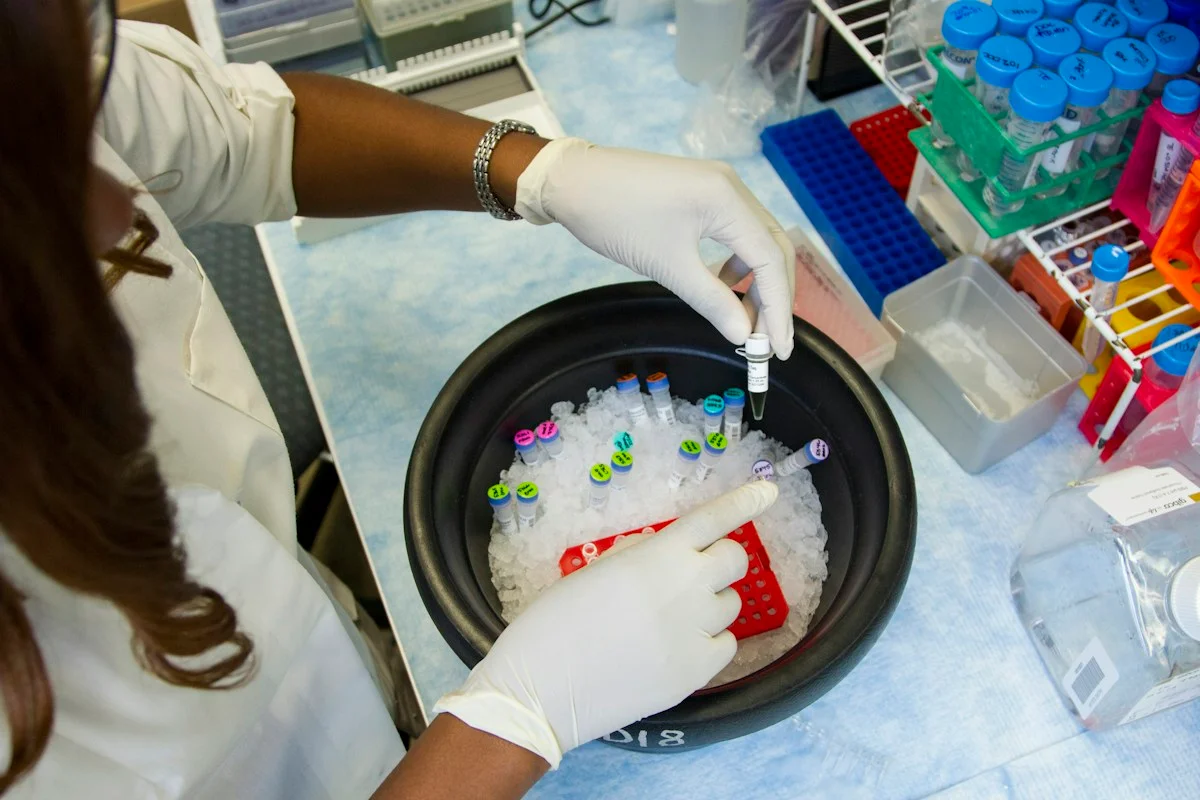
Illustrative photo · not a video frame
The Royal Society · 44 min 28 sec · Background explainer
Is this the AI future we want? | The Royal Society
YouTube loads only when you choose; playback starts when you press play.
A Royal Society discussion with Madhumita Murgia and biomedical engineer Alison Noble on AI’s potential in healthcare, science and culture, recorded at the 30 June 2026 event.
Evidence check: A discussion of opportunities and risks, including future possibilities. It is not evidence that every application described is available or clinically proven.
Published 2026-07-01. Source checked 14 October 2026. Check captions on the publisher’s video page.
Check the healthcare evidencePhoto: Daniel Sone / National Cancer Institute / Unsplash · Licence
Evidence check
Retrospective Chinese cohorts; 84.1% male and 82.5% hepatitis-B positive; residual confounding; inconsistent treatment-line data; locally assessed progression; unpaired CT and MRI; no prospective test of added clinical value.
Reporting basis: Source analysis. Original source date: 17 October 2026.
Explore the full source and evidence record
What this means for people
- Patients could eventually gain more prognostic information from scans they already undergo, but premature use could wrongly change a cancer treatment. The study measured associations, not better treatment choices or quality of life.
Global context
The cohort reflects Chinese liver-cancer practice, where hepatitis B is a dominant cause. Aetiology, body composition, treatment access and imaging protocols differ elsewhere, so the measurements require regional validation.
What the evidence does not yet show
- Retrospective Chinese cohorts; 84.1% male and 82.5% hepatitis-B positive; residual confounding; inconsistent treatment-line data; locally assessed progression; unpaired CT and MRI; no prospective test of added clinical value.
What to watch next
- Prospective multinational validation, paired scan-method comparisons and decision-impact studies showing whether organ features add useful information to established liver-cancer prognostic tools.
Living evidence record
Impact record IAI-06OVFQP
Evidence stage
Studied
Confidence
Supported
Reporting basis
Source analysis
Independent or research support
Present
Record status
Monitoring
Last checked
18 October 2026
Source trail
1 direct source across 1 source type.
People impact
Documented in this record.
Uncertainty
Limits and next checks are explicit.
Stages describe the evidence available—not whether a technology is good or bad. See the public method.
Single-source reporting disclosure
This record analyses one direct source. It can establish what npj Precision Oncology published or reported, but it is not independent corroboration of every performance claim or predicted outcome. The confidence label will change only when broader evidence is added.
Evidence trail
Sources used for this report
Links checked 18 October 2026
Research question, topics and regions
Whether automatically measured abdominal-organ features in pretreatment scans are associated with survival among people with hepatocellular carcinoma receiving immune-checkpoint therapy
Topics: Cancer care · Medical imaging · Immunotherapy · Prognosis · Clinical evidence
Regions: China · Asia · Global
This report is labelled source analysis. We summarise and analyse source material in our own words; company statements remain attributed claims until independently supported. Translated summaries preserve the meaning of the original source and link back to it. Read our editorial standards.
The Impact Brief · Free
Keep following the stories that matter to you.
Get our weekday briefing on what changed and why it matters. Choose your topics below.
Reader commentary
What does this mean where you are?
Tell us what you have seen at work, in your community or in everyday life. A question is welcome too. No account is needed; we review notes before publishing them.
Explore commentary across the portal →Published reader notes
0No published reader notes yet. You can start the evidence-led discussion above.
Prefer a private correction or response? Contact the newsroom.