Can clinical context improve breast-ultrasound AI? A 256-patient test is not decisive
Three hybrid systems divided the honours on one Polish ultrasound dataset. Clinical metadata helped one model find more malignant lesions and draw stronger masks, but another classified cases more accurately—and most differences were not statistically significant.
The short version
3 key points
- What changed
- The study used 256 scans from 256 patients, keeping each patient in only one fold during five-fold cross-validation.
- Why it matters
- Breast-ultrasound AI could eventually support radiologists by highlighting suspicious lesions and drawing boundaries, but false negatives, calibration and workflow effects matter more to patients than a benchmark leader board. This study did not measure those real-world outcomes.
- What to watch
- External, multi-centre validation with locked models and patient-level separation
No single model won every clinically relevant measure
A peer-reviewed study from Vellore Institute of Technology compared three hybrid systems that both classify breast lesions and outline them on ultrasound. The simplest headline is a split decision. A DoubleHeadUNet with a ResNet-34 backbone achieved the highest classification accuracy, 82.94%, and the highest area under the receiver-operating curve, 87.70%. A transformer model conditioned with clinical metadata achieved the highest malignant-lesion recall, 81.63%, and the strongest segmentation scores: Dice 0.901 and intersection-over-union 0.825.[1]
Who it affects
Breast-ultrasound AI could eventually support radiologists by highlighting suspicious lesions and drawing boundaries, but false negatives, calibration and workflow effects matter more to patients than a benchmark leader board. This study did not measure those real-world outcomes.[1][2]
Keep in mind
The evidence is retrospective and restricted to 256 patient scans from one public Polish collection.
There was no external hospital cohort, prospective use, clinician comparison or patient-outcome measurement.
Most accuracy differences between the three models were not statistically significant.
Source analysis · 2 linked sources. Original source date: 18 October 2026.
Read the full report · 4 min The evidence, methods, implications and limitations
No single model won every clinically relevant measure
A peer-reviewed study from Vellore Institute of Technology compared three hybrid systems that both classify breast lesions and outline them on ultrasound. The simplest headline is a split decision. A DoubleHeadUNet with a ResNet-34 backbone achieved the highest classification accuracy, 82.94%, and the highest area under the receiver-operating curve, 87.70%. A transformer model conditioned with clinical metadata achieved the highest malignant-lesion recall, 81.63%, and the strongest segmentation scores: Dice 0.901 and intersection-over-union 0.825.
Those measures answer different questions. Accuracy counts correct labels overall; AUC measures how well a model separates classes across thresholds; malignant recall asks how many malignant cases it catches; Dice and intersection-over-union measure overlap between a predicted lesion outline and the reference mask. Better performance on one does not guarantee better performance on the others. The authors' statistical analysis found that most accuracy differences between the three systems were not significant, so the results do not establish a universally superior architecture.[1]
The patient-level design reduces leakage, but the evidence remains small and internal
The comparison used Breast-Lesions-USG, a public collection of 256 ultrasound scans from 256 adult women examined in Poland between 2019 and 2022. The archive contains 266 segmented benign and malignant lesions plus patient, image and tumour labels; outcomes were confirmed through follow-up care or biopsy. The researchers used five-fold cross-validation with strict patient separation and nested hyperparameter selection. That matters because putting images or lesions from the same person into both training and testing can make a medical-imaging system appear more general than it is.
All results still come from repeated partitions of one relatively small collection. The paper reports no independent hospital, scanner or country held out for external validation, no prospective workflow study and no comparison with radiologists on the same cases. Its strongest numbers therefore describe performance inside this benchmark, not diagnostic accuracy in a clinic. The journal has published the paper as a peer-reviewed, citable early accepted version that may still receive editorial corrections before its final Version of Record.[1][2]
The distinction that matters
What each model led
The three architectures were evaluated on the same patient-separated five-fold procedure, but the leaders changed with the outcome.
ResNet-34 DoubleHeadUNet
Classification accuracy 82.94%
Highest AUC: 87.70%
Swin V2 + U-Net
Classification accuracy 79.76%
AUC: 87.54%
Transformer-FiLM U-Net
Malignant recall 81.63%
Dice 0.901; IoU 0.825
Clinical metadata may be useful, but it must be tested without shortcuts
The metadata-conditioned model is the most practically interesting part of the study. It combines image features with structured clinical information through feature-wise linear modulation, allowing non-image context to alter the network's internal representation. Its higher malignant recall and segmentation overlap suggest that context may help when the task prioritises finding suspicious lesions and tracing their boundaries. But the lower overall classification lead—and the mostly non-significant differences—means that benefit is not yet decisive.
For patients and clinicians, the immediate implication is restraint. This is research on retrospective images, not a screening replacement, treatment recommendation or evidence that AI improves outcomes. A stronger assessment would freeze the models and test them across multiple hospitals, ultrasound devices and patient groups; report calibration, false negatives and performance by age and lesion type; compare clinicians with and without assistance; and measure whether assistance changes biopsies, missed cancers, workload or time to diagnosis. Until then, the study is evidence about promising design choices, not readiness for independent clinical use.[1][2]
Watch the background, then check the evidence
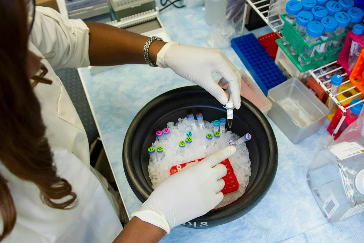
Illustrative photo · not a video frame
The Royal Society · 44 min 28 sec · Background explainer
Is this the AI future we want? | The Royal Society
YouTube loads only when you choose; playback starts when you press play.
A Royal Society discussion with Madhumita Murgia and biomedical engineer Alison Noble on AI’s potential in healthcare, science and culture, recorded at the 30 June 2026 event.
Evidence check: A discussion of opportunities and risks, including future possibilities. It is not evidence that every application described is available or clinically proven.
Published 2026-07-01. Source checked 14 October 2026. Check captions on the publisher’s video page.
Check the healthcare evidencePhoto: Daniel Sone / National Cancer Institute / Unsplash · Licence
Evidence check
The evidence is retrospective and restricted to 256 patient scans from one public Polish collection.
There was no external hospital cohort, prospective use, clinician comparison or patient-outcome measurement.
Most accuracy differences between the three models were not statistically significant.
Reporting basis: Source analysis. Original source date: 18 October 2026.
Continue the story
Read next
Also about medical imaging
Can AI spot liposarcoma on ultrasound?
A peer-reviewed Chinese study reports strong results in a 95-patient internal test, including 0.97 accuracy. But all 317 patients came from one hospital and there was no external validation, so this is a proof of concept—not a clinically cleared diagnostic system.
Read the 8 min reportAlso about medical imaging
Can AI support breast-ultrasound decisions across countries?
A peer-reviewed South Korean-led study validated an interpretable retrieval-augmented system across 8,311 images from 11 cohorts in seven countries and tested assistance with four readers. The retrospective evidence is encouraging, but it is not a prospective screening trial or proof of better patient outcomes.
Read the 7 min reportExplore the full source and evidence record
What this means for people
- Breast-ultrasound AI could eventually support radiologists by highlighting suspicious lesions and drawing boundaries, but false negatives, calibration and workflow effects matter more to patients than a benchmark leader board. This study did not measure those real-world outcomes.
Global context
The models were developed by researchers in India using images collected in Poland. That cross-border reuse is valuable, but it does not establish performance across scanner brands, health systems, ethnic groups or clinical pathways elsewhere.
What the evidence does not yet show
- The evidence is retrospective and restricted to 256 patient scans from one public Polish collection.
- There was no external hospital cohort, prospective use, clinician comparison or patient-outcome measurement.
- Most accuracy differences between the three models were not statistically significant.
What to watch next
- External, multi-centre validation with locked models and patient-level separation
- Calibration and false-negative reporting by lesion type and patient subgroup
- Reader studies comparing radiologists with and without assistance on the same cases
- Prospective evidence about biopsies, missed cancers, workload and time to diagnosis
Living evidence record
Impact record IAI-1MS2XJR
Evidence stage
Studied
Confidence
Supported
Reporting basis
Source analysis
Independent or research support
Present
Record status
Monitoring
Last checked
19 October 2026
Source trail
2 direct sources across 2 source types.
People impact
Documented in this record.
Uncertainty
Limits and next checks are explicit.
Stages describe the evidence available—not whether a technology is good or bad. See the public method.
Related-source reporting disclosure
This record analyses 2 linked source records around the same underlying development. The extra records add method, date or context, but they do not by themselves constitute independent replication of every performance claim or predicted outcome.
Evidence trail
Sources used for this report
Links checked 19 October 2026
Research question, topics and regions
Hybrid AI for breast-ultrasound lesion classification and segmentation
Topics: Breast ultrasound · Medical imaging · Clinical metadata · Cancer diagnosis · Model validation
Regions: India · Poland · Europe · Global
This report is labelled source analysis. We summarise and analyse source material in our own words; company statements remain attributed claims until independently supported. Translated summaries preserve the meaning of the original source and link back to it. Read our editorial standards.
The Impact Brief · Free
Keep following the stories that matter to you.
Get our weekday briefing on what changed and why it matters. Choose your topics below.
Reader commentary
What does this mean where you are?
Tell us what you have seen at work, in your community or in everyday life. A question is welcome too. No account is needed; we review notes before publishing them.
Explore commentary across the portal →Published reader notes
0No published reader notes yet. You can start the evidence-led discussion above.
Prefer a private correction or response? Contact the newsroom.