Can an interpretable AI spot small liver cancers on CT? External testing is promising—but incomplete
A peer-reviewed multicentre study tested dLI-RADS on 133 external patients. Sensitivity reached 93%, versus 78% for radiologist-assigned LI-RADS, but only sensitivity could be compared externally and every lesion was manually segmented.
The short version
- What happened
- The retrospective study used 143 patients for training, 62 for internal evaluation and 133 for external evaluation, representing 451 lesions in total.
- Who it affects
- For people with small liver lesions, a reliable second reader could support earlier, more consistent review. False positives can also trigger anxiety, repeat imaging or invasive work-up, so sensitivity must be weighed against specificity and real clinical consequences.
- What remains unproven
- Retrospective study
Keep in mind
Retrospective study
Manual segmentation
Selected lesions under 40 mm
Predominantly male cohorts
Composite non-cancer reference
External LI-RADS comparison limited to sensitivity
No prospective workflow test
No patient outcomes
Early-access accepted article
Commercial research affiliations
Source analysis · 1 linked source. Primary analysed source date: 30 October 2026.
Keep following the evidence
Join The Impact Brief founding list for selected AI reports, their effect on people and the evidence limits. Read a sample first.
Read the full report · 3 min The evidence, methods, implications and limitations

The separate test hospital is the study’s strongest feature
Researchers combined neural-network predictions with handcrafted imaging measurements that mirror major LI-RADS features, then used logistic regression to estimate whether a manually outlined liver lesion was hepatocellular carcinoma. Training used 143 patients with 196 lesions; internal evaluation used 62 patients with 84 lesions; and a separate external cohort included 133 patients with 171 lesions. Lesions over 40 millimetres were excluded. Histology was the cancer reference, while non-cancer lesions used a composite reference standard.
In the internal set, dLI-RADS reached 80% lesion-level accuracy versus 74% for conventional LI-RADS and met the study’s five-percentage-point non-inferiority test. In the external set, sensitivity was 93% versus 78% for LI-RADS. The model’s external specificity was 74%, overall accuracy 86% and area under the curve 84%. A separate hospital test reduces the risk that the result depends only on one local scanner or workflow.[1]
It is not yet evidence that the system improves diagnosis or care
The external head-to-head comparison is narrower than the headline numbers suggest: LI-RADS features were unavailable for non-cancer lesions, so only sensitivity—not specificity or accuracy—could be compared directly. Every lesion was manually segmented, leaving an important labour and reproducibility step outside the model. The retrospective cohorts were also predominantly men, and the study did not test reporting time, reader behaviour, false-positive work-ups, treatment access or patient outcomes.
Several authors work at Guerbet Research, while the paper reports Région Île-de-France and ANRT funding and declares no competing interests. A prospective multi-reader study should now test automated or routine segmentation, prespecified thresholds, subgroup performance, calibration and downstream decisions. Until then, dLI-RADS is a promising externally tested decision-support method—not a replacement for radiologists or proof of better cancer outcomes.[1]
The distinction that matters
What the three cohorts establish
Separating development from evaluation helps show where the result is strongest and where evidence is still missing.
Training
143 patients; 196 lesions
Built the combined neural-network and imaging-feature model
Internal test
62 patients; 84 lesions
80% accuracy versus 74% for LI-RADS; non-inferiority met
External test
133 patients; 171 lesions
93% sensitivity, but only sensitivity was directly comparable with LI-RADS
Watch the background, then check the evidence
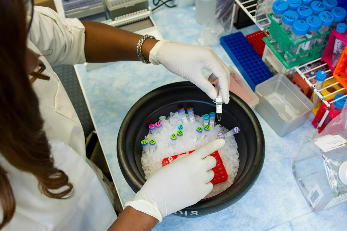
Illustrative photo · not a video frame
The Royal Society · 44 min 28 sec · Background explainer
Is this the AI future we want? | The Royal Society
YouTube loads only when you choose; playback starts when you press play.
A Royal Society discussion with Madhumita Murgia and biomedical engineer Alison Noble on AI’s potential in healthcare, science and culture, recorded at the 30 June 2026 event.
Evidence check: A discussion of opportunities and risks, including future possibilities. It is not evidence that every application described is available or clinically proven.
Published 2026-07-01. Source checked 14 October 2026. Check captions on the publisher’s video page.
Check the healthcare evidencePhoto: Daniel Sone / National Cancer Institute / Unsplash · Licence
Continue the story
Read next
Continue exploring clinical decision support: compare the finding and its evidence limits.
Can interpretable AI distinguish ovarian tumours before surgery?
A peer-reviewed retrospective study trained five classifiers on laboratory data from 349 patients at one Chinese hospital. Its best model reached 93.7% leave-one-out accuracy, but the small reused cohort and absence of external or prospective validation rule out clinical deployment.
Read the next reportExplore the full source and evidence record
What this means for people
- For people with small liver lesions, a reliable second reader could support earlier, more consistent review. False positives can also trigger anxiety, repeat imaging or invasive work-up, so sensitivity must be weighed against specificity and real clinical consequences.
Global context
The development and internal cohorts were assembled in France and the external cohort in South Korea. That cross-country test is useful, but liver-disease causes, scanners, contrast protocols, prevalence and clinical thresholds differ across hospitals and regions.
What the evidence does not yet show
- Retrospective study
- Manual segmentation
- Selected lesions under 40 mm
- Predominantly male cohorts
- Composite non-cancer reference
- External LI-RADS comparison limited to sensitivity
- No prospective workflow test
- No patient outcomes
- Early-access accepted article
- Commercial research affiliations
What to watch next
- Prospective multi-reader evaluation
- Automated segmentation
- Specificity and calibration by subgroup
- Reporting time and workflow effects
- False-positive work-up
- Treatment access and patient outcomes
- Final Version of Record
Living evidence record
Impact record IAI-0VRPU9O
Evidence stage
Studied
Confidence
Supported
Reporting basis
Source analysis
Independent or research support
Present
Record status
Monitoring
Last checked
30 October 2026
Source trail
1 direct source across 1 source type.
People impact
Documented in this record.
Uncertainty
Limits and next checks are explicit.
Stages describe the evidence available—not whether a technology is good or bad. See the public method.
Single-source reporting disclosure
This record analyses one direct source. It can establish what Scientific Reports published or reported, but it is not independent corroboration of every performance claim or predicted outcome. The confidence label will change only when broader evidence is added.
Evidence trail
Sources used for this report
Links checked 30 October 2026
Research question, topics and regions
Whether dLI-RADS can classify small liver lesions on multiphase CT at least as accurately as the conventional LI-RADS assessment
Topics: Liver cancer · Medical imaging · Computed tomography · External validation · Clinical decision support · Interpretable AI
Regions: France · South Korea · Europe · Asia
This report is labelled source analysis. We summarise and analyse source material in our own words; company statements remain attributed claims until independently supported. Translated summaries preserve the meaning of the original source and link back to it. Read our editorial standards.
Reader commentary
What does this mean where you are?
Tell us what you have seen at work, in your community or in everyday life. A question is welcome too. No account is needed; we review notes before publishing them.
Explore commentary across the portal →Published reader notes
0No published reader notes yet. You can start the evidence-led discussion above.
Prefer a private correction or response? Contact the newsroom.