A spinal-cord AI worked across thousands of brain scans—its clinical role remains untested
The multicentre pipeline labelled upper cervical levels and extracted research measurements from routine brain MRI. External validation was large, but the multiple-sclerosis demonstration involved only 51 people and no care decision.
The short version
- What happened
- Training used brain MRI from 998 controls and 879 people with hereditary ataxias across 32 sites; external validation used 2,737 scans from four public datasets.
- Who it affects
- People with neurodegenerative disease may eventually benefit if existing brain MRI can supply reliable spinal-cord measurements without extra scanning, but no change in diagnosis, treatment or outcome was tested.
- What remains unproven
- The longitudinal clinical demonstration included only 51 people with multiple sclerosis
Keep in mind
The longitudinal clinical demonstration included only 51 people with multiple sclerosis
No diagnosis, prognosis or treatment decision was evaluated
Performance was not above 92% in one of five external datasets
Extensive consortium and industry relationships require transparent independent replication
Source analysis · 1 linked source. Primary analysed source date: 23 October 2026.
Keep following the evidence
Join The Impact Brief founding list for selected AI reports, their effect on people and the evidence limits. Read a sample first.
Read the full report · 3 min The evidence, methods, implications and limitations

The pipeline turns a brain scan into a repeatable spinal-cord measurement
A large international team trained three segmentation approaches to find and label the C1-C3 levels of the upper cervical spinal cord in ordinary T1-weighted brain MRI. The development data included 998 controls and 879 people with hereditary ataxias from 32 sites, aged 5 to 85. A separate image classifier was trained to flag labelling errors, while the pipeline also calculates basic image-quality measures.
The best model, nnU-Net, recorded Dice overlap scores of 0.85 at C1 and 0.89 at C2 and C3. The quality-control classifier reported 97.9% accuracy and 98.7% sensitivity. Across 2,737 external scans from four public sources, labelling accuracy exceeded 92% in four of five evaluated datasets. These results support a scalable research tool across heterogeneous scanners and populations.[1]
Detecting change in 51 people is a feasibility result
The team applied the measurement pipeline to longitudinal scans from 51 people with multiple sclerosis and detected upper-cervical atrophy over time. That is evidence the tool can recover a plausible research signal; it is not evidence that the measurement predicts progression, changes treatment or improves care.
A next-stage assessment should lock the pipeline before testing, report failures by scanner and patient group, compare automated measurements with expert review and evaluate a pre-specified clinical question. Public and charitable agencies funded the work, while companies supported the TRACK-FA consortium. Several investigators disclosed industry relationships unrelated to the manuscript; the model developers reported no competing interests.[1]
The distinction that matters
The evidence ladder
The study moves from multicentre development to external validation and a small longitudinal demonstration.
Development
1,877 people across 32 sites
Train C1-C3 labelling and quality control
External validation
2,737 public-dataset scans
>92% accuracy in four of five datasets
MS demonstration
51 people followed over time
Atrophy signal, not clinical utility
Watch the background, then check the evidence
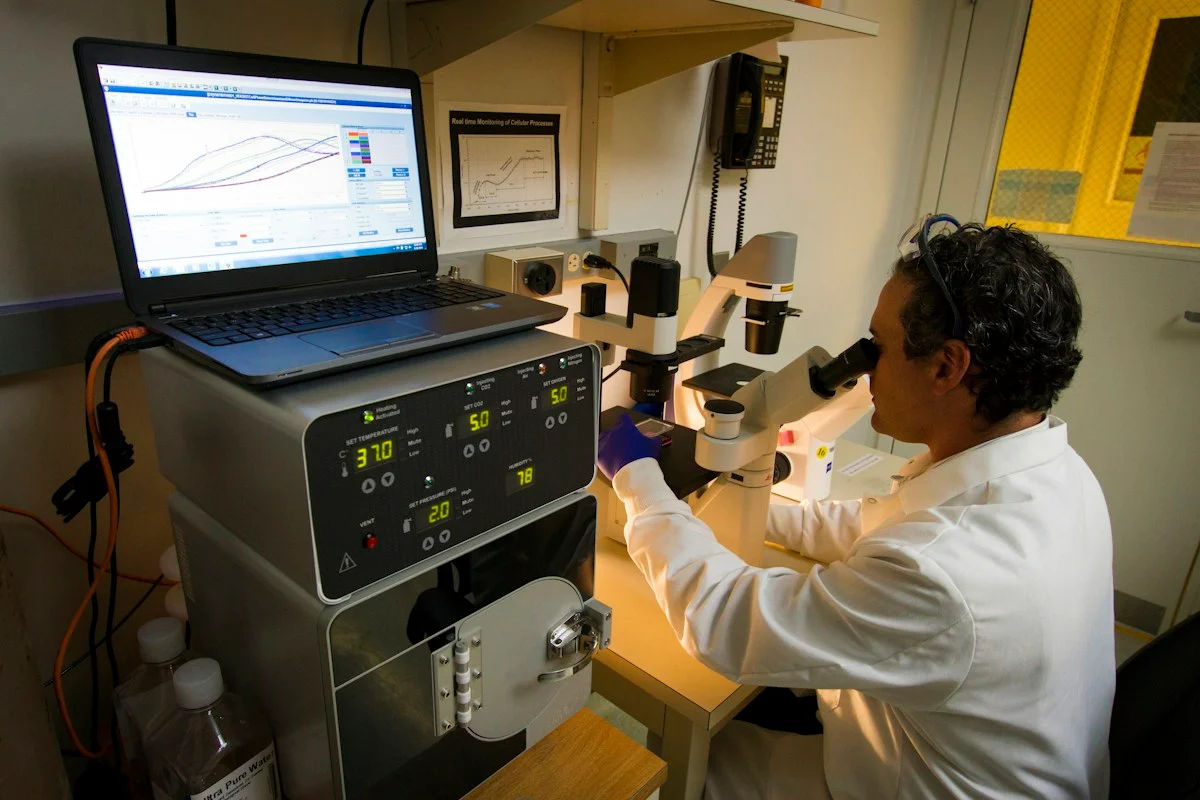
Illustrative photo · not a video frame
Google DeepMind · 1 min 52 sec · Background explainer
Protein folding explained
YouTube loads only when you choose; playback starts when you press play.
A short introduction to why a protein’s shape matters. It provides background for understanding what AI structure prediction contributes to biological research.
Evidence check: This is the developer’s explanation. Predicting a structure does not by itself establish a treatment’s safety or benefit for patients.
Published 2020-11-30. Source checked 14 October 2026. Check captions on the publisher’s video page.
Explore current researchContinue the story
Read next
Continue exploring medical imaging: compare the finding and its evidence limits.
Ear-disease AI narrowed the trainee–expert gap—but also changed 46 correct answers to wrong ones
Thirteen clinicians re-read 100 disease-enriched ear images with AI advice. Accuracy rose most for junior residents, but the single-centre retrospective study cannot show how the tool performs in routine care.
Read the next reportExplore the full source and evidence record
What this means for people
- People with neurodegenerative disease may eventually benefit if existing brain MRI can supply reliable spinal-cord measurements without extra scanning, but no change in diagnosis, treatment or outcome was tested.
Global context
Development involved 32 sites and external public datasets, improving geographic and scanner diversity. Translation into care still depends on local workflow, scanner and population validation.
What the evidence does not yet show
- The longitudinal clinical demonstration included only 51 people with multiple sclerosis
- No diagnosis, prognosis or treatment decision was evaluated
- Performance was not above 92% in one of five external datasets
- Extensive consortium and industry relationships require transparent independent replication
What to watch next
- Locked independent validation
- Failure rates by scanner and demographic group
- Expert-review workload
- Prospective prognosis or treatment-impact studies
Living evidence record
Impact record IAI-1AIJEJA
Evidence stage
Studied
Confidence
Supported
Reporting basis
Source analysis
Independent or research support
Present
Record status
Monitoring
Last checked
25 October 2026
Source trail
1 direct source across 1 source type.
People impact
Documented in this record.
Uncertainty
Limits and next checks are explicit.
Stages describe the evidence available—not whether a technology is good or bad. See the public method.
Single-source reporting disclosure
This record analyses one direct source. It can establish what npj Digital Medicine published or reported, but it is not independent corroboration of every performance claim or predicted outcome. The confidence label will change only when broader evidence is added.
Evidence trail
Sources used for this report
Links checked 25 October 2026
Research question, topics and regions
Whether deep learning can reliably label C1-C3 spinal levels and extract upper-cervical morphometry from heterogeneous brain MRI
Topics: Medical imaging · Spinal cord · Neurodegeneration · Research automation
Regions: Brazil · Australia · Europe · North America · Global
This report is labelled source analysis. We summarise and analyse source material in our own words; company statements remain attributed claims until independently supported. Translated summaries preserve the meaning of the original source and link back to it. Read our editorial standards.
Reader commentary
What does this mean where you are?
Tell us what you have seen at work, in your community or in everyday life. A question is welcome too. No account is needed; we review notes before publishing them.
Explore commentary across the portal →Published reader notes
0No published reader notes yet. You can start the evidence-led discussion above.
Prefer a private correction or response? Contact the newsroom.